
|
|
Chapter 7:
The Cervical Spine
From R. C. Schafer, DC, PhD, FICC's best-selling book:
“Clinical Biomechanics: Musculoskeletal Actions and Reactions”
Second Edition ~ Wiliams & Wilkins
The following materials are provided as a service to our profession. There is no charge for individuals to copy and file these materials. However, they cannot be sold or used in any group or commercial venture without written permission from ACAPress.
All of Dr. Schafer's books are now available on CDs, with all proceeds being donated
to chiropractic research. Please review the complete list of available books.Kinesiology of the Neck Evaluating Gross Muscle Strength of the Neck Evaluating Gross Joint Motion of the Neck General Aspects of Cervical Trauma Injury Incidence Basic Posttraumatic Roentgenographic Considerations of the Neck Classic Effects of Severe Cervical Trauma Soft-Tissue Injuries of the Posterolateral Neck Clinical Biomechanics of the Upper Cervical Spine Regional Structural Characteristics Kinematics of the Upper Cervical Spine Upper Cervical Trauma Clinical Biomechanics of the Lower Cervical Spine Kinematics of the Lower Cervical Spine Lower Cervical Trauma Selected Clinical Problems of the Cervical Spine Cervical Subluxation Syndromes Neurovascular Compression Syndromes Clinical Compression Tests Visual Subluxation Patterns Postural Realignment Traumatic Brachial Plexus Traction Structural Fixations Cervical Disc Disorders Cervical Spondylosis Cervical Scoliosis Torticollis The Troublesome Fifth Cervical Area Rheumatic Disease of the Cervical Spine Ankylosing Spondylitis of the Cervical Spine Cervical Deformities and AnomaliesChapter 7: The Cervical Spine
This chapter considers those factors that are of biomechanical and related clinical interest imperative to the satisfactory evaluation of common or not infrequent cervical syndromes. The discussion assumes that the physician is skilled in taking a thorough clinical history and performing the basic physical, orthopedic, neurologic, and roentgenographic examination procedures. The kinesiology and kinematics of the neck, the effects and mechanisms of cervical trauma, and a number of clinical problems are discussed that are pertinent to the diagnosis and management of musculoskeletal cervical disorders.
Background
The viscera of the neck serve as a channel for vital vessels and nerves, the trachea, esophagus, spinal cord, and as a site for lymph and endocrine glands. The cervical spine provides musculoskeletal stability and support for the cranium, and a flexible and protective column for movement, balance adaptation, and housing of the spinal cord and vertebral artery. When the head is in balance, a line drawn through the nasal spine and the superior border of the external auditory meatus will be perpendicular to the ground.
Cervical subluxations may be reflected in total body habitus, and insults can manifest themselves throughout the motor, sensory, and autonomic nervous systems. Many peripheral nerve symptoms in the shoulder, arm, and hand will find their origin in the cervical spine. Nowhere in the spine is the relationship between the osseous structures and the surrounding neurologic and vascular beds as intimate or subject to disturbance as it is in the cervical region.
Many of the skeletal landmarks readily observed in the thin individual are frequently obscured in the obese (Fig. 7.1). Except for the manatee and some sloths, all mammals have seven cervical vertebrae.
Kinesiology of the NeckThe cervical spine is a miracle in design and structure as it moves in various planes. It must support the head, and it must move the eyes and the ears for various sensory orientations.
Mechanically, the head teeters on the atlanto-occipital joints, shaped like cupped palms tipped slightly medially. Because the line of gravity falls anterior to these articulations, a force must be constantly provided in the upright posture by the posterior neck muscles to hold the head erect. Added to this gravitational stress is the action of the anterior muscles of the neck, essentially the masticatory, suprahyoid, and infrahyoid groups, which as a chain join the anterior cranium to the shoulder girdle.
Flexion, extension, rotation, lateral flexion, and circumduction are the basic movements of the cervical region. Movements of the head on the neck are generally confined to the occiput-atlas-axis complex and can be described separately from movements of the neck on the trunk. The prime movers and accessories involved in neck motion are listed in Table 7.1.
Table 7.1. Neck MotionJoint Motion Prime Movers Accessories Flexion Sternocleidomastoid Scalenes Longus colli Hyoid muscles Longus capitis Rectus capitis anterior Rectus capitis lateralis Extension Trapezius, upper Transversospinalis group Splenius capitis Levator scapulae Splenius cervicis Semispinalis capitis Semispinalis cervicis Erector spinae capitis Erector spinae cervicis Rotation Sternocleidomastoid Scalenes Trapezius, upper Transversospinalis group Spenius capitis Spenius cervicis Lateral Scalenes Transversospinalis group Flexion Levator scapulae Rectus capitis lateralisCervical motions are usually tested with the patient seated unless the patient is unable to hold his head erect. Passive motion should never be attempted if spinal fracture, dislocation, advanced arteriosclerosis, or severe instability is suspected.
Evaluating Gross Muscle Strength of the Neck
Muscle strength is recorded as from 5 to 0 or in a percentage and compared bilaterally whenever possible. Grading has been previously described. The major muscles of the neck, their primary function, and their innervation are listed in Table 7.2.
Table 7.2. Major Muscles of the Neck
  Muscle                     Major Function       Spinal Segment
  Erector spinae, upper      Extension, rotation      C1–T1
  Longus colli               Flexion                  C2–C6
  Longus capitis             Flexion                  C1–C3
  Rectus capitis anterior    Flexion                  C1–C2
  Rectus capitis lateral     Flexion                  C1–C2
  Scalenes                   Flexion, rotation        C4–C8
  Semispinalis capitis       Extension, rotation      C1–T1
  Semispinalis cervicis      Extension, rotation      C1–T1
  Splenius capitis           Extension, rotation      C1–C8
  Splenius cervicis          Extension, rotation      C1–C8
  Sternocleidomastoid        Flexion, rotation        C2, XI
  Trapezius, upper           Extension, rotation      C3–C4
FLEXION
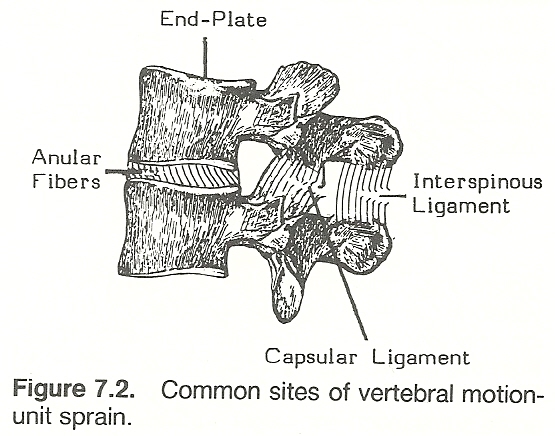 |
Flexion of the neck as a whole is conducted primarily by the sternocleidomastoideus, the longus group, and the rectus capitis anterior and lateralis,
with secondary assistance from the scalenes (Fig. 7.2) and hyoid muscles. Extension is controlled by the upper trapezius, splenius group, the semispinalis
group, and the erector spinae, forming the paravertebral extensor mass. Secondary assistance is provided by several small intrinsic neck muscles and the
levator scapulae.
The position to test strength of the cervical flexors is taken by stabilizing the patient's sternum with one hand to prevent thoracic flexion and
placing the palm of the other hand against the patient's forehead. Strength is
evaluated by having the patient slowly attempt to flex his neck against this
resistance.
EXTENSION
 |
The strength of the many extensors is evaluated by placing the stabilizing
hand in the patient's upper dorsal area to prevent thoracic extension and the
palm of the resisting hand over the occiput of the patient. Strength is measured
by having the patient slowly extend his neck against this resistance. The stabilizing hand may be placed on the superior aspect of the trapezius between the neck and the humerus to palpate muscle contraction at the same time.
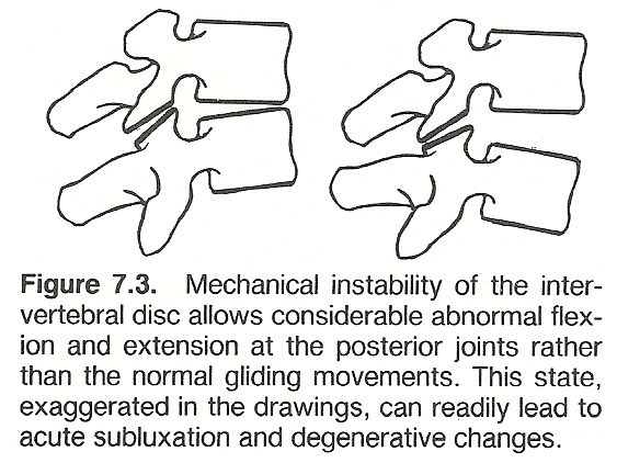
Phillips points out the necessity of normally lax ligaments at the atlantoaxial joints to allow for normal articular gliding, thus making tonic muscle
action the only means by which head stability is maintained. Goodheart feels
that the splenius (Fig. 7.3) is responsible for maintaining head level more than
any other muscle. "Occipital sideslip and jamming frequently are associated
here."
ROTATION
The primary muscles involved in cervical rotation are the sternocleidomastoideus, upper trapezius, and splenius group, with some assistance provided by
the scalenes and intrinsics.
Muscle strength of the cervical rotators is tested by standing in front of
the patient and placing the stabilizing hand on the patient's left shoulder and
the resisting palm against the patient's right cheek when right rotation is
being measured. The examiner's hand positions are switched for testing left rotation strength. Rotational strength is evaluated by having the patient attempt
to slowly rotate his head against the resistance for each side.
LATERAL FLEXION
 |
Lateral flexion is accomplished by the scalenus anticus, medius, posticus,
and the levator scapulae (Fig. 7.4). Secondary assistance is provided by the
small lateral intrinsic muscles of the neck.
Muscle strength of the lateral flexors is tested by standing at the side of
the patient and placing the stabilizing hand on the patient's shoulder to prevent thoracic movement and the resisting palm on the patient's skull above the
ear. Muscle strength is evaluated by having the patient slowly flex his neck
laterally against the resistance.
Evaluating Gross Joint Motion of the Neck
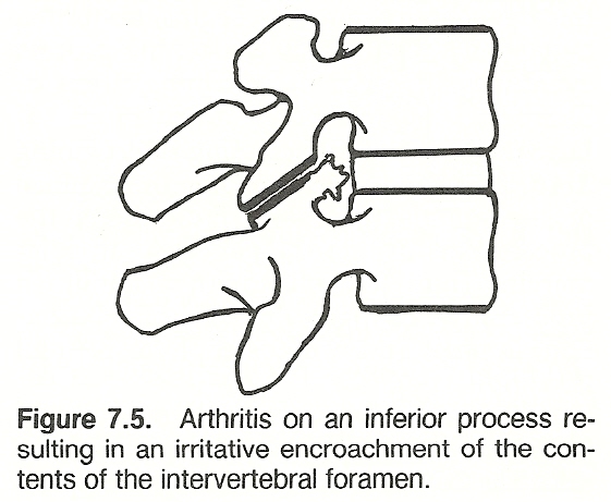
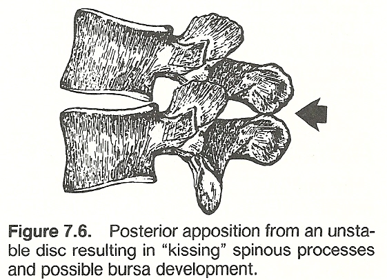
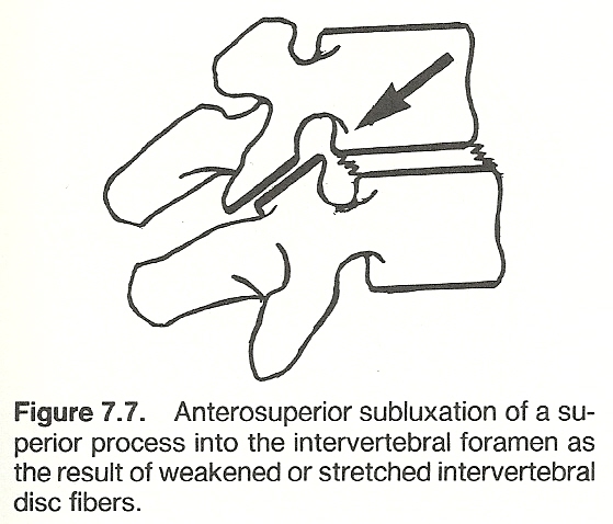
Gross joint motion is roughly screened by inspection during active motions
(Figs. 7.5, 7.6, 7.7). When a record is helpful, it is usually measured by
goniometry. The patient is placed in the neutral position, with the goniometer
centered with its base on line with the superior border of the larynx and the
goniometer arm along the mastoid process. The neutral reading, flexion, extension, rotation, and lateral flexion are recorded.
FLEXION AND EXTENSION
 |
The patient flexes his head as far forward as possible, keeping the goniometer arm along the mastoid process. The end of flexion motion is recorded.
Then, starting from the neutral position, the patient extends his head as far
back as possible, keeping the goniometer arm along the mastoid process. The end
of neck extension is recorded. In cases of ankylosis, the goniometer is placed
to measure the neutral position, and the deviation from this point is recorded.
ROTATION
The patient is placed in the neutral position, and the patient's shoulders
are steadied with the hands. The patient rotates his head as far to the right
and left as possible. The arc of motion is estimated eparately for right and
left motion by the position of the patient's chin in relation to his shoulder.
The goniometer is not necessary for this evaluation. In situations of ankylosis,
the angle at which the cervical region is fixed is estimated by noting the position of the patient's chin and the angle is recorded.
LATERAL FLEXION
 |
The patient is placed in the neutral position with his arms abducted to
steady the shoulders. The goniometer is centered over the back of the neck with
the base on the C7 spinous process, and the goniometer arm is extended along the
midline of the neck. The neutral reading is recorded. Then the reading is
recorded after the patient has bent his neck as far to the left as possible, the
reading being taken after the end of lateral flexion. The reading for the right
side is recorded in the same manner. In cases of ankylosis, deviations are
recorded from the neutral position.
Blows to the head or neck may result in unconsciousness, but most blows do
not. Rather, the effect is a "subconcussive" or "punch drunk" effect for a few
moments. This state may be the effect of a severe blow to the head or the cumulative effects of many blows. It is assumed that the reader is well acquainted
with the proper emergency procedures involved in head and neck trauma.
(1) mild (eg, contusions, strains);
Neurapraxia. Recovery of nerve contusion usually occurs within 6 weeks.
Nerve contusion may be the result of either a single blow or through persistent
compression. Fractures and blunt trauma are often associated with nerve contusion and crush. Peripheral nerve contusions exhibit early symptoms when produced
by falls or blows. Late symptoms arise from pressure by callus, scars, or supports. Mild cases produce pain, tingling, and numbness, with some degree of
paresthesia. Moderate cases manifest these same symptoms with some degree of
motor and/or sensory paralysis and atrophy4.
Incidence. Strains (Grades 1–3) or indirect muscle injuries are common,
frequently involving the erectors. Flexion and extension cervical sprains are
also common (Grades 1–3) and usually involve the anterior or posterior longitudinal ligaments, but the capsular ligaments may be involved. In the neck
especially, strain and sprain may co-exist. Severity varies considerably from
mild to dangerous. The C1 and C2 nerves are especially vulnerable because they
do not enjoy the protection of an IVF.
Mechanisms. Other than those in automobile accidents, the forces in whiplash
are usually administered from below upward; eg, an uppercut blow to the chin or
a blow to the forehead while running forward. This is in contrast to the compressive type of hyperextension or hyperflexion injury where the force is usually from above downward. Thus, knowing the direction of force, even if the magnitude is unknown, is important in analyzing the effects. A facial injury usually
suggests an accompanying extension injury of the cervical spine as the head is
forced backward.
Mechanisms. An occipital injury usually suggests an accompanying flexion injury of the anterior cervical spine and posterior soft tissues as the skull is
forced forward (Fig. 7.10). Flexion injury may also be a part of whiplash,
superimposed upon an extension injury.
Therapy. Peripheral inhibitory afferent impulses can be generated to partially close the pre-synaptic gate by acupressure, acupuncture, or transcutaneous nerve stimulation. Most authorities feel deep sustained manual pressure on
trigger points is the best method, but a few others prefer almost brutal short-duration pressure (1–2 seconds). Deep pressure is contraindicated in any patient
receiving anti-inflammatory drugs (eg, cortisone) as subcutaneous hemorrhage may
result.
Common Sites
Reference patterns vary considerably according to the severity and chronicity
of the trigger point phenomenon involved.
Passive Stretch. Mild passive stretch is an excellent method of reducing
spasm in the long muscles. Heavy passive stretch, however, destroys the beneficial reflexes. One technique, for example, is to place the patient prone on an
adjusting table in which the headpiece has been slightly lowered. The patient's
head is turned toward the side of the spastic muscle. With head weight alone
serving as the stretching tensile force, the spasm should relax within 2–3 minutes. Thumb pressure, placed on a trigger area, is then directed toward the
muscle's attachment and held for a few moments until relaxation is complete.
For study, it is best to divide the cervical spine into upper and lower
regions because of its anatomic design and functional arrangement. The upper
spine is composed of the occipital condyles, the atlas, and the axis. It is different morphologically and functionally from the lower cervical spine that is
made up of vertebrae C3–C7. The axis is thus a transitional vertebra in that
its superior aspect is part of the upper complex and its inferior aspect is part
of the lower complex.
The Lateral Masses and Articular Processes. The lateral masses are oval in
shape with an oblique anteromedial articulation. They must support the weight of
the head, which comprises about 7% of body weight. The articular surfaces on the
superior side of the lateral masses are biconcave to allow for the seating and
movement of the biconvex occipital condyles and are relatively large to dissipate the weight of the head. The inferior articulating facets of the atlas have
an inferomedial articulation with anteroposterior convexity to fit the articulation of the superior facets of the axis. These inferior facets of the atlas lie
directly underneath the superior facets, unlike those of subjacent apophyseal
joints whose inferior facets lie posterior to the superior facets.
The Anterior Arch. The C1–C2 joint is an unusual joint in that the inner
anterior arch of C1 has a small facet that is in contact with the odontoid process of C2, separated only by a small synovial cavity. A small synovial bursa
also separates the posterior aspect of the odontoid from the cruciate ligament
(Fig. 7.17). The osseous and ligament complex of this area allows great rotation and some flexion and extension. The anterior arch of C1 normally remains 1
mm from the odontoid in flexion and extension. If there is widening of this
space greater than 3 mm in the adult or 4 mm in the child, damage to the transverse ligament of the atlas can be suspected. Note: The specific range of cervical motion differs quite widely among so many authorities that any range offered here should be considered hypothetical
depending on individual planes of articulation, other variances in structural
design (eg, congenital, aging degeneration, posttraumatic), and soft-tissue
integrity. This wide variance in opinion is also true for the centers of motion
described.
Flexion Range. Without any neck participation, the head can be moved 10° in
flexion between the occiput and atlas, according to Cailliet (Fig. 7.22). During
strict upper neck flexion, the condyles roll backward and slide slightly posterior on the atlas while the atlas rolls anteriorly and somewhat superiorly,
taking the odontoid with it so that the dens slightly approaches the clivus of
the basiocciput. As the atlas slides anteriorly in relation to the condyles, the
posterior arch of the atlas and occiput separate only slightly, but this is
exaggerated if movement is virtually isolated at the atlanto-occipital joint
(eg, ankylosing spondylitis). Also during flexion, the inferior lateral masses
of the atlas roll upward posteriorly and slide backward on the superior facets
of the axis for about 5°. Opening of the superior aspect of the atlanto-odontoid
space is not appreciably restricted by the delicate cruciate ligament. Movement
is restricted mainly by the apophyseal capsules, the ligamentum flavum, the
interspinous ligament, the posterior nuchal muscles, and impact of the chin
against the sternum.
Range. During rotation, the occipital condyles and the atlas initially move
as one unit on the axis (Fig. 7.23). Approaching the end of the range of motion,
the condyles can rotate several degrees (8°–10°) upon the atlas in the direction of movement. Only a few authorities contest this fact. C1 rotation occurs
about the dens of C2 which serves as a pivot. As mentioned, 50% of total neck
rotation occurs between C1 and C2 (capable of 80°–100° rotation) before any
rotation is noted from C2 to C7 or at the atlantal-occipital joint. After about
30° of atlas rotation on the dens, the body of the axis begins to rotate,
followed by progressively diminishing rotation in the remaining cervical segments. Because the atlantal-occipital and atlantoaxial apophyseal articulations
are not horizontal, rotation must be accompanied by a degree of coupled tilting.
If a complete fixation occurs between C1 and C2, the remaining cervical
segments tend to become hypermobile in compensation. Thus, gross inspection of
neck rotation (or other motions) should never be used to evaluate the function
of individual segments.
Range. Normally, about a 45° tilt can be observed between the skull and the
shoulder. About 5° of this occurs at the atlanto-occipital joint (Fig. 7.24),
following the arc of the condyles on the superior facets of the atlas; and 6°
occurs at the atlantoaxial joint, following the arc of the inferior facets of
the atlas on the superior facets of the axis. As the occiput and atlas shift
laterally as one unit towards the concavity during lateral bending, the space
between the dens and lateral mass of the atlas widens on the concave side. At
the same time, the occipital condyles translate slightly laterally on the
superior facets of the atlas toward the convexity and the atlas slips slightly
toward the side of concavity. These movements are quite slight unless there is
a degree of instability involved. If the atlanto-occipital capsular ligaments
are weakened, the condyle on the side of lateral bending may strike the tip of
the odontoid. The body of the axis tends to rotate towards the concavity while
its spinous process shifts toward the convexity due to the coupling mechanism.
Flexion/Extension. The prime mover of atlanto-occipital flexion is the rectus
capitis anterior, aided by the longus capitis. The range is limited essentially
by the elasticity of the posterior ligaments and by the tip of the dens meeting
the bursa below the anterior rim of the foramen magnum. Extension is powered by
the rectus capitis posterior group. Extension and lateral tilt of the upper
cervical region is restricted by tension of the tectorial membrane and the
posterior arch of the atlas becoming trapped between the occiput and the axis.
Flexion/Extension. In addition to the rolling motion of the atlas on the
occiput, the atlas is capable of some tilting where the anterior ring of the
atlas moves upward on the odontoid and the posterior arch rides downward, or
vice versa (Fig. 7.25). During severe flexion, there could be considerable
separation of the anterior arch of the atlas from the odontoid, but it is checked by the weak transverse arms of the cruciate and by tension of the stronger
tectorial membrane. Extension is more readily resisted when the anterior arch
meets the odontoid and the interarticular tissues compress.
Disturbances in this area usually arise from muscular spasm of one or more
of the six muscle bundles that have attachments on the occiput, atlas, or axis.
Unequal tension and ultimate fibrotic changes within the paravertebral structures can readily influence the delicate nerve fibers and vascular flow. The
vertebral artery is frequently involved by compression of the overlying muscles
in the suboccipital triangle. In fact, West points out that the vertebral artery
has been completely occluded by turning the head backward and to the opposite
side during postmortem studies. Even without a degree of arteriosclerosis, the
vertebral artery can be considered a quite firm tube in the adult that responds
poorly to twisting and pressure.
Right/Left Condyle Inferior or Superior. A unilateral suboccipital muscle
spasm causes the affected condyle to be pulled deep into the articulating concavity of the atlantal lateral mass on one side (sunken condyle). This may not
be attended by a degree of rotation. Inspection from the back shows a low,
medially inclined mastoid process on the side of involvement (Fig. 7.26). Palpation discloses the mastoid riding close to the transverse process of the atlas,
tension and tenderness in the groove between the mastoid and the lower jaw, and
fullness in the groove between the occiput and the posterior ring of the atlas
on the side of involvement. A right or left condyle superior may be considered
the converse aspect of a right or left condyle inferior. That is, as one condyle
is pulled inferior and anterior, the other condyle presents a superior and
posterior picture, or vice versa. There are certain situations, however, that
indicate a unilateral abnormality without converse adaptation. This latter condition usually follows a blow to the vertex downward when the head is somewhat
laterally flexed and the condyle on the side of the concavity is jammed into the
lateral mass of the atlas (eg, spearing tackle).
(1) inspection from the back shows the head held in a stiff inferior position with some
posterior deviation; and
Suboccipital Jamming. This common subluxation, usually of a trigeminal
(ophthalmic division) reflex nature, is often seen in people under severe visual
or mental stress. Irritative impulses cause contraction of suboccipital muscles
that pull the occiput upon the posterior arch of the atlas, creating a painful
bilateral condylar jamming (Fig. 7.27). A compressive vertex blow is a rare
cause. Palpation reveals suboccipital spasm, tenderness, nodular swellings, and
a closing of the inferior nuchal ridge on the posterior arch of the atlas.
Although the condition is usually bilateral, one side may be affected more than
the other.
Right or Left Lateral Atlas. An atlantal sideslip between the atlas and axis
articulations is usually attended by a degree of superiority and anteriority on
the side of laterality because of the inclination of the articulating surfaces.
Only in cases of severe twisting trauma will this not be the case. Ipsilaterally, palpation will reveal the transverse process of the atlas to be more lateral
and slightly superior and anterior than its counterpart.
A clinical test, suggested by Goodheart, is to have the patient lying
supine, then passively rotating the head right and left. If an anterior atlas
subluxation exists on the left, the atlas has already turned to the right so
that the patient's head will turn much further to the right. But when it is
turned from right to left, the atlas cannot come out of its fixed anterior position on the left, thus motion is relatively restricted. This test is a valid
indication only in the absence of muscular spasm or some other type of motion
barrier restricting rotation (eg, lower cervical unilateral fixation).
Classes. The atlas may be fractured at its posterior arch, ring, or anterior
arch. There are six common types of severe injury, all of which are serious.
Keep in mind that nontraumatic dislocations of the upper cervical complex are
more common than traumatic dislocations (eg, congenital anomalies, arthritis,
infection), and their possibility should never be overlooked. Atlanto-occipital dislocation. This usually, but not always, anterior
displacement of the occiput on the atlas occurs from a severe horizontal force
from behind that shears the skull across the atlas, rupturing the articular capsules, and damaging the medulla. This rare occurrence can often be accurately
evaluated by computing Powers ratio on a lateral roentgenograph (Fig. 7.31).
Atlas dislocation with fractured dens. The atlas may displace anteriorly
on the axis or the occiput posteriorly on the atlas and fracture the odontoid
process if the ligaments hold. The force may be hyperextensive or hyperflexive.
The patient may survive if extreme care is taken in transportation to the hospital. If the transverse ligament is avulsed from the atlas, a small fragment of
bone may lie between the odontoid and the cord. If the odontoid is displaced
posteriorly, the situation is usually fatal because of injury to the cord. Posttraumatic spontaneous fusion of C1 to the occiput is always a potential complication if the patient survives.
Fractured posterior arch of the atlas. This usually occurs from a severe
vertical compression force during extension where the lateral masses are fixed
between the condyles and the pillars of the axis and the posterior ring fractures and displaces outward. A base fracture of the odontoid is often associated. If a fracture line is not evident on lateral roentgenography (differentiated from congenital clefts), headache, suboccipital pain, stiffness, acute
suboccipital jamming, and subtle signs of basilar insufficiency from compression
of the vertebral artery should still stimulate suspicions.
Of all atlantoid fractures, most literature states that those of the posterior arch are the most common yet easily overlooked as the displacement is usually mild. The common site is at the narrowest portion just posterior to each
lateral mass, usually at the groove for the vertebral artery. Retropharygeal
swelling is usually absent, and oblique views are often necessary for demonstration.
Jefferson fracture. A more severe vertical compression blow may split the
atlas and burst the lateral masses outward, disrupting both the anterior and
posterior rings into several fragments (Fig. 7.32). Ring fractures are frequently produced by blows on top of the head where vertical forces are dispersed laterally. Keep in mind that if a severe axial force is produced through the skull
downward, the inclined condyles of the occiput serve as a mechanical wedge upon
the atlas. This is usually evident in an open-mouth x-ray view. Overhang of the
atlantal lateral masses and widening of the paraodontoid space will be associated. Severity depends upon fragment displacement relative to the cord and other
vital tissues. That is, if the ligaments do not retain these fragments, death
from cord damage will be likely. Atlas-axis displacement. In C1–C2 A–P dislocations, C1 most often displaces anteriorly relative to C2. If a force comes from the back, undoubtedly the
muscles will be unprepared and the force will meet minimum resistance. Yet,
anterior dislocation is rare, and posterior displacement is even more infrequently seen. Forward dislocation widens the predental space and alters a
roentgenographic line connecting the cortices of the anterior parts of the spinous processes from C1 to C7, unless the process of C2 is fused or congenitally
short. If this is suspected, careful flexion-extension views or a C1–C2 tomogram
is recommended. The mechanism of injury is usually hyperflexion or hyperextension; and even in moderate cases, signs of trauma to the occipital nerve should
be evident. In rare instances where there are sufficient traction forces to
rupture the anterior longitudinal ligament, the anterior ring of the atlas may
be lifted up and over the dens so that an intact odontoid is seen anterior to
the anterior ring.
Orthopedic rotary subluxation of the atlas on the axis. Forced rotation
of the upper neck may produce a locked rotary displacement of a lateral mass of
the atlas on the subjacent superior facet of the axis. This requires atlantal
rotation in excess of 45° on the axis. A neurologic deficit is not commonly
involved. The patient will appear with his head rotated to one side and cocked
away from the side of rotation ("cock robin" position). Care must be taken to
differentiate this sign which is also so common in acute torticollis.
Types: The classic order of Anderson/D'Alonzo is applicable:
Type I: Avulsion of the upper part of the odontoid. This is rare.
Nature has made many structural adaptations in the cervical region because
of the small structures, the required range of motion, and the enlarged cord in
this region as compared to other spinal regions (Fig. 7.34). The laminae are
slender and overlap, and this shingling increases with age. The osseous elevations on the posterolateral aspect that form the uncovertebral pseudojoints tend
to protect the spinal canal from lateral IVD herniation, but hypertrophy of
these joints added to IVD degeneration can readily lead to IVF encroachment.
Segmental Angulation. Angulation of one vertebral segment on a lateral
roentgenograph in excess of 11° greater than an adjacent vertebra that is not
chronically compressed is also indicative of instability and pathologic displacement (Fig. 7.39). While conservative traction may reduce the associated
displacement, it is doubtful that a normal resting position can be guaranteed
without surgical fusion in severe cases.
As opposed to the primary thoracic kyphosis which is a structural curve, the
cervical and lumbar anterior curves are functional arcs produced by their wedge-shaped IVD's and they normally flatten in the nonweightbearing supine position.
Likewise, they quickly adapt to changes involving the direction of force.
Incidence. Cervical kyphosis occurs most frequently after the age of 40, and
the sexes appear equally affected. The cause is often the result of trauma-producing whiplash injury, herniated disc, subluxation, dislocation, fracture,
and/or ligamentous (especially posterior) injury. Torticollis, arthritis, malignancy, tuberculosis, osteomyelitis, and other pathologies may be involved.
Compression Injuries. Vertebral body crush fractures are rare, and less common in the cervical spine than elsewhere (Fig. 7.43). They are the result of a
vertical force, often during flexion, such as that of a football "spearing
tackle". Compression fractures of articular processes occur in extension injuries to the neck. They are not common with the exception of those occurring from
automobile "whiplash" injuries and diving into shallow water. They are not
usually demonstrable on A–P or lateral films until deformity is severe, but
oblique views will often demonstrate them. They are best seen on "pillar" views.
The pillar view is taken with the trunk A–P and the head turned 45° to the side.
These views, to be taken bilaterally, will show the articular pillar in profile.
Apophyseal fractures are frequently quite apparent when present in pillar views.
(1) the dens will fracture and displace laterally;
Rotary Injuries. These are often found combined with flexion, extension, and
lateral flexion injuries. Keep in mind that while the cervical ligaments are
quite resistant to pure flexion and extension stress, they are far less resistant to shear stress (Fig. 7.47). It is for this reason that:
(1) the anterior
longitudinal ligament is often torn when the neck is overextended and rotated
and
(1) lesser occipital nerve neuralgia,
involving the posterior area of the occipitofrontalis muscle, mastoid process,
and upper posterior aspect of the auricle;
De Rusha suggests that dysphagia and dysarthria may at times be due to upper
cervical involvement rather than a central nervous system situation. The C1
joins the hypoglossal cranial nerve which supplies the intrinsic muscles of the
tongue. It then descends to join the descending cervical which is derived from
C2 and C3. A loop of nerves, the ansi hypoglossi, which supplies muscles
necessary for deglutition and speaking, is derived from C1–C3.
Sensory Changes. When direct nerve root involvement occurs on the posterior
root of a specific neuromere, it manifests as an increase or decrease in sensitivity over the dermatome. A typical example includes foraminal occlusion or
irritating factors exhibited clinically as hyperesthesia, particularly on the
dorsal and lateral aspects of the thumb and radial side of the hand, when involvement occurs between C5–C6. Another example is on the dorsum of the hand,
the index and middle fingers, and the ventroradial side of the forearm, thumb,
index and middle fingers, when involvement occurs between C6–C7. In other
instances, this nerve root involvement may cause hypertonicity and the sensation
of deep pain in the musculature supplied by the neuromere. For example, in C6
involvement, there is deep pain in the biceps; or in C7 involvement, there is
deep pain in the triceps and supinators of the forearm. Direct pressure near the
nerve root or along its distribution may be particularly painful.
Loss of disc space, especially in the lower cervical area, may contribute as
a source of chronic irritation to an already inflamed root by altering the angulation of the IVF tunnel. The sequence of inflammation, granulation, fibrosis,
adhesion formation, and nerve root stricture may follow, along with a loss in
root mobility and elasticity. These degenerative changes are not as pronounced
during youth.
(1) suboccipital or postocular migraine;
Phillips states that if a subluxation produces a stretching of the paravertebral musculature, there will be a continuous barrage of afferent impulses in
the Group Ia fibers. "These afferent impulses monosynaptically bombard the alpha
motor neurons causing the paravertebral musculature to go into tetany. There is
a cessation of this afferent barrage when the stretch is released. The muscle
stretching also initiates afferent impulses in the Group II afferents from
flower spray endings which may reinforce the spastic muscle condition." He goes
on to say that trauma to facet joints, disturbed articular relationships, spasms
of closely related muscles, and overlying trigger points –all the result of a
subluxation– set up a barrage of flexor-reflex afferent impulses via the Group
II–IV fibers that converge upon the internuncial pool in lamina 7 of the spinal
cord. "This abundant supply of flexor-reflex afferent impulses excites the alpha
motor neurons through multisynaptic connections causing an excess of excitation
of paravertebral muscles resulting in spasm."
The Vagus Nerve. As the vagus lies almost in immediate contact with the
transverse process of the atlas, rotary subluxation of the atlas may cause pressure which produces a wide range of symptoms. The syndrome produced may exhibit
as nasal and sinus congestion, swallowing and speech difficulties, cardiac
arrythmias, functional coronary artery spasm, gastric and intestinal colic, and
other symptoms of vagal disturbance.
The Cervicothoracic Junction Area. The area of cervicothoracic transition is
a complex of prevertebral and postvertebral fascia and ligaments subject to
shortening. It offers a multitude of attaching and crossing muscles such as the
longus colli, trapezius, scaleni, sternocleidomastoid, erector spinae, interspinous and intertransverse, multifidi and rotatores, splenius capitis, splenius
cervicis, semispinalis capitis, semispinalis cervicis, longissimus capitis,
longissimus cervicis, and the levator costarum and scapula –all of which are
subject to spastic shortening and fibrotic changes that tether normal motion.
Differential diagnosis must exclude a cervical rib etiology from infectious
neuritis, banding adhesions, arthritis of the shoulder joint, clavicle fracture
callus, bifid clavicle, cervical arthritis, subacromial bursitis, 1st rib
subluxation and posttraumatic deformities, spinal or shoulder girdle malignacies, Pancoast's tumor of the lung apex, and cardiac disease. Aneurysms of the
subclavian artery are rare.
Differentiation of Neural vs Circulatory Symptoms. Compression of nerve
tissue results in numbness, pain, paralysis, and loss of function. Compression
of vascular structures results in moderate pain and swelling. The obstruction of
circulation can result in clotting within the vessels with possible consequent
infarction in the tissues supplied. These unilateral phenomena are fairly
limited to the cervicobrachial distribution. When the arm is depressed, the clavicle moves inferior and anterior, and
this widens the space between the clavicle and 1st rib.
When the shoulder is depressed, the upper and middle trunks of the
brachial plexus are stretched tightly over the tendinous edge of the scalenus
medius and the lower trunks are pulled into the angle formed by the 1st rib and
the scalenus medius tendon. Most symptoms found on shoulder depression will be
the result of this traction. There is no compression of the subclavian artery
against either scaleni.
When the shoulder is retracted, the clavicle does not impinge upon the
subclavian vein but the tendon of the subclavius muscle compresses the vein
against the 1st rib. The middle third of the clavicle pushes the neurovascular
bundle against the anterior scalenus medius, and this could cause compression if
a space-occupying lesion is also present (eg, cervical rib, extrafascial band).
(1) manipulation of the head on the neck can produce coordinated flexion and extension movements
on the paralyzed arm and leg of a hemiplegic patient,
Resisted Flexion. The patient sits erect with the head straight. The patient
is instructed to place clasped hands against the forehead, breathe normally, and
attempt to push the head forward against the resisting hand contact (Fig. 7.58). This position should be held for about 7 seconds; then the patient relaxes for
several seconds and repeats the exercise a few times.
NERVE PINCH OR STRETCH SYNDROMES
Ipsilateral vs Contralateral Symptoms. If the symptoms appear on the opposite side of the forceful bending, undoubtedly a nerve has been "pinched" within
the powerful muscles dorsal to the sternocleidomastoid. If this is the case, the
symptoms usually subside in a few minutes with only slight residual tenderness
and paresthesia, which disappears within a few hours. On the other hand, if
symptoms appear on the same side as the direction of the forceful bending, deep
skeletal injury such as severe rotary subluxation, fracture, dislocation, or
nerve compression may be involved4.
(1) flaccid, atrophic paralysis of the muscles supplied by the involved nerve
and
In ulnar nerve damage, sensation is lost on the medial side of the hand,
including the little finger and medial half of the ring finger. In median nerve
damage, sensation of the remainder of the anterior surface of the hand is lost.
However, motor involvement is the main feature as sensory loss is often obscured
by overlapping innervation. As time goes by after severe nerve injury, the
affected part assumes a posture and atrophy peculiar to the particular nerve
involved; for example, "wrist drop" with the radial nerve, "claw hand" with the
ulnar nerve, "flat hand" with the median nerve, and "ape hand" with the ulnar
and median nerves.
(1) normal and abnormal
segmental motion and
During motion palpation, each cervical vertebra is palpated during flexion,
extension, rotation, and lateral flexion to assess segmental mobility. The
amount of motion in any particular joint depends upon:
(1) the shape of the joint
surface,
Atlanto-occipital Palpation. Spinal motion palpation usually starts with the
articulation between the occiput and the atlas. Flexion, extension, rotation,
and lateral flexion should be evaluated. Flexion-extension. In cervical extension, the atlas projects forward as a
unit; in flexion, the atlas rolls backward. The examiner's palpating finger is
placed within the space between the tip of the transverse of the atlas and the
ramus of the jaw, while the supporting hand on the patient's scalp forces the
patient's head into extension so that the chin moves up and forward and then
into flexion so that the chin moves down and inward. Note the change in space
underneath the palpating fingertip. The space should normally open on extension
and close on flexion. These movements, conducted several times, should be
restricted as much as possible to the upper cervical area and tested bilaterally.
Rotation. In rotating the atlas horizontally anterior without flexion or
extension, there is normally a wider separation between the jaw and the transverse process. That is, the transverse-jaw space opens as the head is turned
away from the palpating finger and closes as the head moves towards the palpating finger. This is a difficult motion to palpate because of the bulging of the
sternocleidomastoideus.
Lateral flexion. The tip of the palpating finger is placed deep within
the small space between the tip of the C1 transverse process and the mastoid
process of the occiput. As the support hand rocks the patient's scalp laterally,
the space change should be noted as the ipsilateral occipital condyle rolls
laterally up and out on the atlas as the scalp moves away from the palpating
finger and down and in as the head is laterally flexed toward the palpating
finger. On flexion to the right, the transverse-mastoid space should open on the
left and close on the right.
Differentiating Atlanto-occipital Muscular and Ligamentous Fixations. The
tip of the palpating finger is placed under the posterior occiput, midway between the occipital notch and the mastoid process. Some examiners prefer to cup
the atlas in the web of the palpating hand so that the thumb palpates one side
while the first finger palpates the other side. The supporting hand rocks the
patient's head into flexion and extension. If a stubborn ligamentous fixation is
present, the fibrous tissues will palpate as a hard mass that does not change
texture during motion. Flexion-extension. The palpating finger is placed in the space between
the posterior tubercle of the atlas and the spinous of the axis. This space
should open on flexion and close on extension, and the posterior tubercle should
become more apparent on flexion and be lost to touch on extension.
Rotation. The tips of the first three fingers are placed horizontally in
the suboccipital space so that the first finger firmly presses against the
occipital notch and the third finger rests lightly on the tip of the C2 spinous
process. The free hand is used to rotate the head. During rotation, several
degrees of atlas rotation should take place before the axis begins to move.
Normally, the third finger will slip upon the spinous process of the axis as the
head is rotated because the head moves 1 cm or more prior to axial motion.
Bilateral atlantoaxial fixation is indicated if the axis immediately follows the
movement of the head (essentially the atlas), noted by the third finger not
gliding over the process of the axis. If unilateral (pivotal) fixation is present, this situation will occur during rotation to one side but not to the
other, and the center of movement will be at the point of fixation rather than
at the odontoid. If the axis is fixed unilaterally, rotary movement will also be
felt on the free side during A–P motion.
Lateral flexion. It has been Gillet's experience that abnormal lateral
flexion of the atlas on the axis is affected most by hypertonicity of the intertansversarii and/or the upper aspect of the longus coli. Motion restricted can
be determined by placing the tip of the palpating finger in the posterolateral
space between the transverse processes of the atlas and axis. Space changes are
checked during both lateral flexion and A–P motion. While intertransversarii
hypertonicity restricts lateral bending, a small degree of lateral gliding of
the atlas on the axis is usually allowed. This does not appear to be true when
hypertonus of the longus coli exists. Flexion-extension. In flexion and extension, the interspinous spaces
should be felt to open and close. Rotation. Areas of rotary fixation are quite easily determined except in
the athlete with extremely heavy posterior neck muscles. The patient rotates the
head as far as possible in one direction. Then the palpating fingers slide
down the posterolateral aspect of the transverse processes. The contact is made
by the tips of the palpating fingers pushing the belly of the sternocleidomastoideus anterior. A firm "bulge" will be evident over the restricted transverse,
and this is usually attributed to a hypertonus multifidus or intertransversarii
muscle. Lateral flexion. The axis is palpated in lateral bending as moving away
from the flexion. To evaluate lateral gliding of the axis, the examiner's thumb
is firmly pressed against the posterolateral aspect of the C2 spinous process,
while the supporting hand moves the patient's scalp in wide lateral flexion.
The third cervical may be palpated during lateral bending, flexion, and
extension much like the axis, noting the separation and closure of the spinous
process on A–P motion and rotation and lateral bending by palpating the posterolateral space between the transverse processes. Rotation reveals minimum motion
and is difficult to palpate. The same procedure is applied to the rest of the
cervical spine. However, palpation in the middle and lower cervical region is
difficult because the palpating finger is usually against tender nerves. Some
examiners prefer an interlaminae contact. Interspinous fixation. Hypertonicity of one or more extensors tends to
bind spinous processes together so that a local lordosis is formed. This condition, often found at the C3–C4 level, is palpable when the spinous processes
refuse to open during forced flexion. It is also often evident on lateral
flexion roentgenographs where two or more vertebrae do not follow the curve of
the neck as a whole.
Covertebral articular fixation. Fixation is common at the lips of the
joints of Luschka by longus colli hypertonicity, ligamentous shortening, and
exostosis. Restricted motion can often be determined during A–P motion from the
anterolateral by carefully pushing the esophagus lateral with two palpating
fingers and evaluating the motion of the vertebral bodies. If during passive
extension it is found that the patient's neck stops sharply at a point far short
of normal extension, Gillet refers to this "brick wall" sign of strong restriction as an indication of cervical osteophytes. This is a classic sign of chronic
degeneration found in the cervical joints of the elderly presenting a thin,
"dry" cervical spine. These fixations usually produce a chronic brachialgia.
Common Sites of Muscular Fixation. There are six major pairs of anterior and
posterior muscles operating in the atlanto-occipital area to produce A–P rocking
of the occiput on the atlas and atlas rotation. Any one or more of these muscles
can be in a state of hypertonicity, thus maintaining the numerous types of upper
cervical subluxation. Obliquus capitis superior. This muscle joins the transverse process of
the atlas to the occiput. Hypertonicity restricts contralateral extension and
lateral flexion (Fig. 7.65).
Obliquus capitis inferior. This muscle spans between the spinous process
of the axis and the transverse process of the atlas. Hypertonicity will tend to
fixate the atlas-axis articulation, especially on rotation toward the opposite
side. The spinous process of the axis will often be palpated as being distinctly
pulled laterally.
Rectus capitis posterior (minor and major). The minor arises from the
posterior tubercle of the atlas and the major from the spinous process of the
axis. Both insert at the occiput and function in extension of the head upon the
neck. Hypertonicity produces approximation of the C2 spinous process and the
occiput, thus increasing upper cervical lordosis and restricting flexion mobility. Gillet feels this state is usually the manifestation of a lower fixation
(eg, anterior thoracic).
Rectus capitis anterior and lateralis. The anterior part originates on
the lateral mass of the atlas and inserts in the basilar part of the occiput.
The lateral aspect arises from the transverse process of the atlas and inserts
at the jugular process of the occiput (See Fig. 7.9). Both serve to flex and
support the head. Hypertonicity restricts extension.
Longus colli. Hypertonicity of the cervical branches of this muscle produces a greater fixed space between the C2 spinous process and the occiput. The
picture is the converse of rectus capitis posterior shortening.
Longus capitis. This muscle arises from the transverse processes of the
C3–C6 vertebrae and inserts at the basilar portion of the occiput. It functions
in head flexion.
The A–P Prime Movers. Hypertonicity of the sternocleidomastoideus (Fig.
7.66) and related flexors forces increased mobility upon the posterior arches, a
decrease in height of the anterior anulus, and restricts extension. The disorder
may be either unilateral or bilateral. This is the converse of generalized
posterior cervical muscle shortening that restricts flexion, decreases posterior
IVD thickness, and forces increased mobility upon the anterior anular fibers.
Either anterior or posterior hypertonicity tends to decrease the normal range of
rotation but less so than hypertonicity of the rotatores (Fig. 7.67).
The Anterior Longitudinal Ligament. This appears to be the only frequent
site of adverse ligamentous fixation in the lower cervical region (See Fig.
7.21).
C2 disc protrusion (C3 nerve root level): posterior neck numbness and pain
radiating to the mastoid and ear. The reflexes test normal.
Vertebral Artery Compression. Associated subluxation and osteophyte development may produce vertebral artery compression, especially if a degree of arteriosclerosis is present (See Fig. 6.12). Symptoms of unsteadiness, dizziness, and
fainting spells will occur especially when the head is rotated to the opposite
side.
(1) conservative treatment fails to produce remission of symptoms;
(1) flattening of the cervical spine from muscular spasm and adhesion development,
The Davis series may suffice, but special views, tomography, myelography, or
discography may be necessary for firm diagnosis.
Severe Trauma. Traumatic dislocations of upper cervical vertebrae cause a
distortion of the neck much like that of torticollis. A rotary fracture-dislocation of a cervical vertebra, especially of the atlas on the axis or the
axis on C3, will produce neck rigidity and a fast pulse, but fever is absent.
Local and remote trigger points are frequently involved. Even in mildly suspicious cases, the neck should always be x-rayed in two or more planes before it
is physically examined.
Barge states that the structural cause of torticollis is a rotatory vertebral malposition and abnormal disc wedging, where the nucleus of an involved
disc has been forced to shift away from compressive forces. The patient's symptoms are often self-limiting with time and rest that allows the disc to expand
in its nonweightbearing (decompressed) state and the vertebral facets to be
relieved of their jammed position. It can be theorized, however, that if the
neck does not achieve this subluxation correction through disc imbibition a
rotatory scoliosis is produced in adaptation so that the victim may at least
have a straight eye level. But, as the now chronic subluxation has not been
fully corrected, it can serve as a focus for morbid neurologic and degenerative
processes, especially at the zygapophyses, covertebral joints, and IVF's.
Type I: Lateral Torticollis. The patient's neck is rigidly flexed laterally
and locked, and usually accompanied by a degree of rotation of the chin away
from the side of tilt. The spinous process of the involved vertebra will often
palpate as being distinctly lateral as compared to its neighbor above and below. Splenius capitis. Increased tone tends to pull the C5–T3 spinous processes lateral, superior, and anterior and to subluxate the occiput inferior,
medial, and posterior.
Scalenus anterior. Hypertonicity tends to pull the C3–C6 transverse
processes inferior, lateral, and anterior and the 1st rib superior and medial.
Scalenus medius. Excessive tone tends to pull the C1–C7 transverse processes inferior, lateral, and anterior and the 1st rib superior and medial.
Scalenus posterior. Hypertonicity tends to pull the C4–C6 transverse
processes inferior, lateral, and anterior and the 2nd rib superior and medial.
Obliquus capitis superior. Increased tone tends to roll the occiput
anterior and inferior and pull the atlas posterior and superior to produce a
lateral occiput tilt and condyle jamming.
Obliquus capitis inferior. Increased tone tends to produce a rotary
torque of the atlas-axis motion unit.
Rectus capitis posterior major. Hypertonicity tends to pull the occiput
posterior, inferior, and medial and the spinous of the axis superior, lateral,
and anterior. Strong hypertonicity will lock the occiput and axis together so
that they appear to act as one unit even though they are not contiguous.
Interspinales. Excessive muscle tone between the spinous processes tends
to hyperextend the motion unit.
Sternocleidomastoideus. Increased tone tends to pull the sternum and clavicle posterior and superior and the occiput inferior and anterior.
Upper trapezius. Hypertonicity tends to pull the occiput posteroinferior,
the C7–T5 spinous processes lateral, and the shoulder girdle medial and
superior.
Range of Motion. In the previous chapter, it was mentioned that normal spinal motion in the pure horizontal plane occurs only at the center of the curves
(Fig. 6.9). While 50% of A–P motion of the cervical spine takes place between
the occiput and the atlas, the remainder is distributed among the other cervical
vertebrae with C5 and C6 making the greatest contribution. This is also true in
lateral bending and rotation below the axis. Thus, if C5 becomes fixed, compensatory effects (and symptoms) will be exhibited at the upper cervical and upper
dorsal areas. This is readily confirmed empirically. In abnormal cervical kyphosis, it will be found that C5 is most frequently the center vertebra of the
affected segmental region and symptomatic picture.
The above deformities are described in detail in standard radiologic atlases and
do not require repetition here. However, a few points are worthy of review in
this section.
Congenital Basilar Coarctation and Platybasia. Basilar coarctation is the
state where the tip of the odontoid lies abnormally high above Chamberlain's or
McGregor's line. The disorder should not be confused with platybasia, an anthropometric flattening of the base of the skull as seen in Down's syndrome. This is
often associated with congenital atlantoaxial subluxation, which occurs in 20%
of mongoloid children. Platybasia is measured by the angle extending from the
clivus and the episthion that is greater than 130°. Such development deformity
must be differentiated from basilar impression.
(1) in acquired
fusions, the margins of the vertebral body tend to be irregular, disc lines are
wider than the adjacent vertebral bodies, the posterior arches are frequently
subluxated,
Significance. A cervical rib arising from C7 and ending free or attached to
the T1 rib appears in the neck as an angular fullness which may pulsate owing to
the presence of the subclavian artery above it. It rarely produces symptoms, and
it is often first noticed when percussing the apex of the lung. The bone can be
felt behind the artery by careful palpation in the supraclavicular fossa and
demonstrated by roentgenography. Pain or wasting in the arm and occasionally
thrombosis may occur from impaired circulation.
General Aspects of Cervical Trauma
The anterior and lateral aspects of the neck contain a wide variety of vital
structures that have no bony protection. Partial protection is provided by the
cervical muscles, the mandible, and the shoulder girdle. After spinal injury, a
careful neurologic evaluation must be conducted. Note any signs of impaired
consciousness, inequality of pupils, or nystagmus. Do outstretched arms drift
unilaterally when the eyes are closed? Standard coordination tests such as
finger-to-nose, heel-to-toe, heel-to-knee, and for Romberg's sign should be
conducted, along with superficial and tendon reflex tests. For reference, the
segmental functions of the cervical nerves are listed in Table 7.3.
Cervical spine injuries can be classified as being:
(2) moderate (eg, subluxations, sprains, occult fractures, nerve contusions, neurapraxias);
(3) severe (eg, axonotmesis, dislocation, stable fracture without neurologic deficit); and
(4) dangerous (eg, unstable fracturedislocation, spinal cord or nerve root injury).
Table 7.3. Segmental Function of Cervical Nerves
Segment   Function

  CERVICAL PLEXUS (C1–C4)

  C1      Motor to head and neck extensors, infrahyoid, rectus capitis
          anterior and lateral, and longus capitis.

  C2      Sensory to lateral occiput and submandibular area; motor,
          same as C1 plus longus colli.

  C3      Sensory to lateral occiput and lateral neck, overlapping
          C2 area; motor to head and neck extensors, infrahyoid,
          longus capitus, longus colli, levator scapulae, scaleni,
          and trapezius.

  C4      Sensory to lower lateral neck and medial shoulder area;
          motor to head and neck extensors, longus coli, levator
          scapulae, scaleni, trapezius, and diaphragm.

  BRACHIAL PLEXUS (C5–T1):

  C5      Sensory to clavicle level and lateral arm (axillary
          nerve); motor to  deltoid, biceps; biceps tendon reflex.
          Primary root in shoulder abduction, exits between
          C4–C5 discs.

  C6      Sensory to lateral forearm, thumb, index and half of
          2nd finger (sensory branches of musculocutaneous nerve);
          motor to biceps, wrist extensors; brachioradialis tendon
          reflex. Primary root in wrist extension, exits between
          C5–C6 discs.

  C7      Sensory to second finger; motor to wrist flexors,
          finger extensors, triceps; triceps tendon reflex.
          Primary root in finger extension, exits between
          C6–C7 discs.

  C8      Sensory to medial forearm (medial antebrachial nerve),
          ring and little fingers (ulnar nerve); motor to finger
          flexors, interossei; no reflex applicable. Primary root
          in finger flexion, exits between C7–T1 discs.

  T1      Sensory to medial arm (medial brachial cutaneous nerve);
          motor to interossei; no reflex applicable. Primary root
          in finger abduction, exits between T1–T2 discs.
Injury Incidence
Due to its great mobility and relatively small structures, the cervical
spine is the most frequent site of severe spinal nerve injury and subluxations.
A wide variety of cervical contusions, Grade 1–3 strains and sprains, subluxations, disc syndromes, dislocations, and fractures will be seen as the result of
trauma. The peak incidence of cervical injury occurs in the 3rd decade, with the
vast majority of the accidents occurring in males. Body build does not appear to
be a major factor. High-speed activities have the highest injury rate.
Considerable cervical spine injury can be attributed to the small, curved
vertebral bodies, the wide range of movement in many planes, and the more laterally placed intervertebral articulations which require the nerve roots to leave
the spinal canal in an anterolateral direction. There is greater space within
the cervical canal than below, but this space is occupied by cord enlargement.
The axis and C6 are the most vulnerable to injury according to accident statistics. The atlas is the least involved of all cervical vertebrae. In terms of
segmental structure, the vertebral arch (50%), vertebral body (30%), and IVD
(30%) are most commonly involved in severe cervical trauma. While the anterior
ligaments are only involved in 2% of injuries, the posterior ligaments are involved in 16% of injuries.
Basic Posttraumatic Roentgenographic Considerations of the Neck
A well-founded appreciation of normal variations, epiphyseal architecture,
development defects, and congenital anomalies is a distinct aid in evaluating
injuries of the cervical area. After the age of 8 years, the neck, with few
exceptions, attains an adult form in which growth plates present few diagnostic
problems.
On the standard lateral and A–P views, the anterior and posterior soft
tissues deserve careful inspection. Signs of widened retrotracheal space,
widened retropharyngeal space, displacement of the prevertebral fat stripe,
laryngeal dislocation, or tracheal displacement should be sought. Abnormal vertebral alignment may be exhibited by a loss of the normal lordotic curve or even
an acute kyphotic hyperangulation, vertebral body displacement, abnormal dens
position, widened interspinous space, or rotation of the vertebral bodies.
Abnormal joints may portray unusual IVD-space symmetry or widening of an apophyseal joint space. It is easy to miss lower cervical fractures inasmuch as they
are often obscured on lateral views by the subject's shoulders if proper precautions are not taken.
Classic Effects of Severe Cervical Trauma
COMPRESSION FORCES
Excessive compression forces on the neck commonly lead to facet jamming
and fixation, isolated or multiple fractures of the atlantal ring, or vertical,
oblique, or bursting fractures of the lower cervical bodies.
HYPERFLEXION FORCES
Excessive anterior bending forces may produce hyperflexion sprain of the
posterior ligaments, compressive wedging of the anterior anulus and vertebral
body, anterior subluxation, anterior bilateral or unilateral dislocation with
locked facets, and spinous process avulsion. Abnormal widening of a spinous
interspace on a lateral roentgenograph should arouse suspicion of ruptured
posterior ligaments.
HYPEREXTENSION FORCES
The effects of posterior bending moments may include hyperflexion sprain of
the anterior ligaments, wedging of the posterior anulus and vertebral body,
posterior subluxation, horizontal fracture of the anterior arch of the atlas,
fracture of the anteroinferior margin of a vertebral body, compression of the
posterior arch and associated structures, posterior bilateral or unilateral
dislocation, spinous process fracture, and traumatic spondylolisthesis.
HYPERROTARY FORCES
Excessive segmental rotation about the longitudinal axis produces anterior
or posterior ligament torsion overstress, rotary subluxation, spiral loosening
of the nucleus pulposus, and unilateral or bilateral atlas-axis dislocation.
The traumatic moments involved invariably include shear forces.
SHEAR FORCES
Excessive shearing forces create disruption of the anterior or posterior
ligaments, end-plate displacement, anterior or posterior subluxation or dislocation, anterior or posterior fracture displacement of the dens, and anterior
compressive fracture of the anterior ring of the atlas or a vertebral body.
LATERAL HYPERFLEXION FORCES
The effects of excessive lateral bending include transverse process fracture, uncinate process failure, lateral dislocation-fracture of the odontoid
process, lateral wedging of the anulus and vertebral body, and brachial plexus
trauma.
Soft-Tissue Injuries of the Posterolateral Neck
CERVICAL CONTUSIONS
Contusions in the neck are similar to those of other areas. They often occur
in the cervical muscles or spinous processes. Painful bruising and tender swelling will be found without difficulty, especially if the neck is flexed. They
present little biomechanic significance unless severe scarring occurs.
DIRECT NERVE TRAUMA
Nerve trauma occurs from contusion, crushing, or laceration.
Axonotmesis. After nerve crush, recovery rate is about an inch per month
between the site of trauma and the next innervated muscle. If innervation is
delayed from this schedule or if the distance is more than a few inches, surgical exploration should be considered.
Neurotmesis. Laceration from sharp or penetrating wounds is less frequently
seen than tears from a fractured bone's fragments. Surgery is usually required.
Stretching injury typically features several sites of laceration along the nerve
and is usually limited to the brachial plexus.
GENERAL ASPECTS OF STRAINS AND SPRAINS
Anterior injuries are more common to the head and chest as they project
further anteriorly, but a blunt blow from the front to the head or chest may
result in an indirect extension or flexion injury of the cervical spine. In any
spinal injury, rarely is the trauma the product of a single force. For example,
while extension, flexion, and lateral flexion injuries are often described
separately in this chapter, rotational, compressive, tensile, and shearing forces are invariably part of the picture.
Typical Signs and Symptoms. Cervical sprain and disc rupture are often associated with severe pain and muscle spasm and are more common in adults because
of the reduced elasticity of supporting tissues. Pain is often referred when the
brachial plexus is involved. Cervical stiffness, muscle spasm, spinous process
tenderness, and restricted motion are common. When pain is present, it is often
poorly localized and referred to the occiput, shoulder, between the scapulae,
arm or forearm (lower cervical lesion), and may be accompanied by paresthesiae.
Radicular symptoms are rarely evident unless a herniation is present. Spasm of
the sternocleidomastoideus and trapezius may be due to strain or irritation of
the sensory fibers of the spinal accessory nerve as they exit with the C2–C4
spinal nerves.
Case Management. Diagnosis and treatment are similar to that of any muscle
strain-sprain, but concern must be given to induced subluxations during the initial strain. Palpation will reveal tenderness and spasm of specific muscles. In
acute scalene strain, both tenderness and swelling will usually be found. When
the longissimus capitis or the trapezius are strained, they stand out like stiff
bands.
Prognosis. Many cervical strains heal spontaneously but may leave a degree
of fibrous thickening or trigger points within the injured muscle tissue. Residual joint restriction following acute care is more common in traditional
medical care than under mobilizing chiropractic management.
EXTENSION STRAIN/SPRAIN
The head may be flexed forward so that the chin strikes the sternum or
thrown sidewards so that the ear strikes the shoulder and the neck can still be
within the normal range of motion. It is most rare, however, that the occiput
strikes the back and does not exceed normal cervical extension.
Kinematics. In whiplash resulting from a mild automobile collision, the cervical trauma is due to indirect trauma from acceleration-deceleration forces. If
the head does not strike anything, the injury is produced solely by inertia forces (Fig. 7.8). The body is moving as a whole at the same speed as the automobile. If the automobile is struck from the rear, the unrestrained head is whipped backward because the head is not restrained by the seat, and then rebound
forward. If the automobile is struck from the front or hits a relatively immovable object, the head is thrown forward and then rebound backward. Thus, the
inertia force displaces the head in the direction opposite to the automobile's
acceleration. The first movement is that of translation which produces a shearing force at the base of the neck because the bending moment is greatest at that
point.
The rebound is caused by several factors. In a front-end collision, for
example, there is an initial flexion elongation of the cervical spine after
impact that is followed by a rebound extension. The rebound is produced by the
rapid deceleration of the automobile, the impact from the seat, and the stretch
reflex produced within the stretched neck and upper dorsal muscles. This reflex
can be quite severe, and because it occurs when the neck is at its full range of
movement, the pull generates considerable compression as well as extension.
Effects. When the head is violently thrown backwards (eg, whiplash), the
damage may vary from minor to severe tearing of the anterior and posterior
longitudinal ligaments. This flattens the cervical curve in about 80% of cases,
and a degree of facet injury must exist even if not evident on film. Stretching
to the point of hematoma may occur in the sternocleidomastoideus, longus capitis, longus cervicis, and scalene muscles (Fig. 7.9). Severe cord damage can
occur that is usually attributed to momentary pressure by the dura, ligamentum
flavum, and laminae posteriorly, even without roentgenologic evidence. Even
without any cord deficit, severe damage to the nerve roots may occur as the
facets jam together and close upon the IVF, especially if fracture occurs. Incidence is highest at the C4–C6 area. Severe stretching of the vertebral arteries
and sympathetic trunk to some degree is inevitable.
Cailliet points out that it is difficult to visualize a sprain causing rupture of the ligaments of a joint without causing some derangement of the opposing joint surfaces, which by definition is an orthopedic subluxation. If a
whiplash injury is considered a severe sprain, an orthopedic subluxation injury
must be assumed to have occurred even if it has been spontaneously reduced.
Such subluxations may occur during the initial movement and/or the rebound movement, and it is not unusual to have manifestations of a flexion sprain superimposed upon manifestations of an extension sprain. In the typical whiplash
injury, whether it be from hyperextension or hyperflexion or both, the effects
of traumatic elongation and compression are compounded by underlying fixations,
arteriosclerosis, spondylosis, ankylosing spondylitis, etc.
Case Management. Treatment of mild or moderate injuries not exhibiting
severe neurologic trauma requires reduction of subluxation, physiotherapeutic
remedial aid, a custom-fitted supporting collar for several weeks depending upon
the clinical symptoms and signs, and graduated therapeutic exercises beginning
with isometric contractions. Continuous traction, which reduces the cervical
lordosis, may be helpful in extension injuries after the acute stage, but it
would usually be contraindicated where the cervical curve has reversed (eg,
flexion strain).
FLEXION STRAIN/SPRAIN
Slight anterior subluxation is usually not serious, but neurologic symptoms
may appear locally or extend down the arm.
Effects. The posterior paraspinal tissues are overstretched, the facets are
sprung open, and the process of bleeding, edema, fibrosis, and adhesions is initiated. Fractures of end-plates may be difficult to assess early. Disc degeneration and posttraumatic osteoarthritis may follow, which leads to spondylosis.
Case Management. Management is similar to that of extension injuries except
that the period of necessary immobilization is often shorter (6–8 weeks).
LATERAL FLEXION STRAIN/SPRAIN
Traumatic brachial plexus traction syndromes will be discussed later in this
chapter. These usually occur when the neck is not only severely flexed sideward
but also flexed forward and down so that the head is anterior to the shoulder.
TRIGGER POINTS
The cervical and suprascapular areas of the trapezius, usually a few inches
lateral to C7, frequently refer pain and deep tenderness to the lateral neck
(especially the submastoid area), temple area, and angle of the jaw (Fig. 7.11).
The sternal division of the sternocleidomastoideus refers pain chiefly to the
eyebrow, cheek, tongue, chin, pharynx, throat, and sternum. The clavicular division refers pain mainly to the forehead (bilaterally), back of and/or deep
within the ear, and rarely to the teeth. Other common trigger points involved in
"stiff neck" are in the levator scapulae, the splenius cervicus lateral to the
C4–C6 spinous processes, and the splenius capitis over the C1–C2 laminae (Fig.
7.12). These points are often not found unless the cervical muscles are relaxed
during palpation.
MYOFASCIAL TRIGGER POINTS IN THE NECK AND BACK
Visceral or somatic trigger-point irritation can produce a degree of spasm
of the paravertebral muscles ipsilaterally in 2–3 segments on the same side as
the entering afferent. However, if the irritation is severe, this effect will
spread up, down, and contralateral (eg, as in renal colic). In this regard,
Stoddard reminds us that the sharp "textbook" demarcation made between the somatic and autonomic nervous systems is erroneous.
Although one or more trigger points may occur in any muscle, they usually
form in clusters and certain muscles and muscle groups (eg, the antigravity
muscles) appear to be more liable than others. See Table 7.4
Table 7.4. Common Trigger Point Syndromes*
     UPPER BODY
     Location                     Primary Reference Zone or Symptoms
     Infraspinatus                Posterior and lateral aspects of the shoulder.

     Intercostal muscles          Thoracodynia, especially during inspiration.

     Levator scapulae             Posterior neck, scalp, around the ear.

     Pectoralis major             Anteromedial shoulder, arm.

     Pectoralis minor             Muscle origin or insertion.

     Quadratus lumborum           Anterior abdominal wall, 12th rib, iliac crest.

     Rectus abdominus             Anterior abdominal wall.

     Semispinalis capitis         Headache, facial pain, dizziness.

     Splenius cervicis            Headache, facial pain, dizziness.

     Sternocleidomastoideus       Headache, dizziness, neck pain, ipsilateral ptosis,
                                  lacrimation, conjunctival reddening, earache,
                                  facial and forehead pain.

     Trapezius                    Lower neck and upper thoracic pain, headache.

LOWER BODY
     Location                     Primary Reference Zone or Symptoms
     Anterior tibialis            Anterior leg and posterior ankle.

     Gastrocnemius/soleus         Posterior leg, from popliteal space to heel. These
                                  trigger points may be involved in intermittent
                                  claudication.

     Gluteus medius               Quadratus lumborum, tensor fasciae latae, gluteus
                                  maximus and minimus, sacroiliac joints, hip,
                                  groin, posterior thigh and calf, cervical exten-
                                  sors, upper thoracic muscles.

     Tensor fasciae latae         Lateral aspect of the thigh, from ilium to the knee.
* Adapted from Sola, with slight modification.
ADJUNCTIVE THERAPIES
The reduction of spasm is often necessary prior to structural correction and
to maintain a corrected position after adjustment.
Therapeutic Heat or Cold. Heat is also helpful, but cold and vapocoolant
sprays have shown to be more effective in acute cases.
Therapeutic Exercise. Mild isotonic exercises are useful for improving circulation and inducing the stretch reflex, especially in the cervical extensors.
These exercises should be done supine to reduce exteroceptive influences on the
central nervous system. In chronic cases, relaxation training with biofeedback
is helpful.
Traction. The effects of cervical traction are often dramatic but sometimes
short lived if a herniated disc is involved. Extreme care must be taken in
posttraumatic cases to eliminate the possibility of instability prior to traction. For example, the use of traction following traumatic spondylolisthesis
in which the anterior longitudinal ligament has been separated can produce
severe displacement with catastrophic effects (Fig. 7.13).
In any vertebral, occipital, or pelvic subluxation, physiotherapy, traction,
muscle relaxants, gross manipulations, muscle stretching, injections, or other
methods will not offer much relief by themselves unless the fixated articulation
is correctly adjusted so that intrinsic function can be normalized.
Clinical Biomechanics of the Upper Cervical Spine
Regional Structural Characteristics
The spinal canal of the upper cervical region is relatively large to accommodate the cervical enlargement of the cord. The pedicles, apophyseal joints,
uncinate processes, and transverse processes have characteristics peculiar and
specific to the cervical spine.
THE ATLAS
In several anatomic respects, the atlas can be considered a sesamoid between
the occiput and axis that serves as a biomechanical washer or bearing between
the occipital condyles and the axis (Fig. 7.14). The atlas is an elongated bony
ring with right and left lateral masses, an anterior arch, a posterior arch, and
bilateral transverse processes that extend from each lateral mass. The absent
body of the atlas is represented by its anterior arch and the dens of the axis.
The inner aspect of the anterior arch contains a facet for the dens. An IVD does
not exist between the occiput and the atlas, nor does the atlas exhibit IVF's or
a distinct spinous process.
The Posterior Arch. The posterior arch of the atlas thickens posteriorly to
where it forms the posterior tubercle (Fig. 7.15). The posterior arch is grooved
to offer some bony protection for the vertebral artery which runs just behind
the lateral mass. This groove is a frequent fracture site.
The Transverse Processes. Only the lumbar vertebrae have transverse processes that extend further from the midline than the atlas. This great width increases the leverage of the muscles that insert at the transverse processes. Unlike other cervical vertebrae, the transverse processes of the atlas are not
grooved to allow egress of a nerve root. The transverse processes of the atlas,
as other cervical vertebrae, contain a conduit (foramen transversarium) for the
vertebral artery.
THE AXIS
The inferior facets of the atlas fit the superior facets of the axis like
epaulets on sloping shoulders (Fig. 7.16). The plane is about 110° to the vertical. To allow maximum rotation of the upper cervical complex without stress to
the contents of the vertebral canal, the instantaneous axis of rotation is
placed close to the spinal cord (ie, near the atlanto-odontoid articulation).
Rotational Restriction. Rotation of C2 on C3 is limited by a mechanical
blocking mechanism that protects the vertebral artery from excessive torsion.
The anterior tip of the superior articular process of C3 impinges on the lateral
margin of the foramen transversarium of C2. This same blocking mechanism is also
found in the subjacent cervical vertebrae (Fig. 7.18).
THE OCCIPITOCERVICAL LIGAMENTS
The cross-shaped cruciate ligament completely secures the odontoid process.
Its main portion is the triangular bilateral transverse ligament, which passes
posteriorly on the dens and connects to the lateral masses of the atlas, transversing in front of the spinal cord. Its main function is to restrict anterior
translation of the atlas. There are also two vertical bands. One rides from the
dens up to the basiocciput, and the other extends from the dens posteriorly down
to the body of the axis. Because these ligaments are often tough, the odontoid
will usually fracture prior to ligament failure. In addition, accessory atlantoaxial ligaments extend superiorly and laterally from the base of the inferior
vertical cruciate and join the base of the dens with the inferomedial aspect of
the lateral mass of C1.
Anterior to the upper arm of the cruciate lie the apical and alar ligaments.
The thin, elastic apical ligament connects the tip of the dens to the anterior
margin of the foramen magnum, and the stronger lateral alar ligaments connect
the medial aspect of the occipital condyles obliquely with the superolateral
aspect of the odontoid (Fig. 7.19). These three guy-wire ligaments, collectively
called the dentate ligaments of the dens, tend to limit rotation and lateral
bending, but their capabilities are quite limited.
The atlantoepistrophic ligament runs between the anterior body of the axis
and the inferior aspect of the anterior ring of the atlas, and the atlantooccipital ligament connects the superior aspect of the anterior ring of the
atlas and the occipital tubercle.
The posterior longitudinal ligament terminates upward as the strong, broad,
fan-shaped membrana tectoria which extends superiorly from the base of the odontoid, over the posterior dens, then obliquely angles forward to blend with the
dura and the clivus of the basiocciput periosteum at the anterior aspect of the
foramen magnum. Its most posterior aspect joins the occiput to the posterior
ring of the atlas, and it serves to check excessive A–P motion. Its deep lateral
part connects the posterior body of C2 with the anterolateral ring of the atlas.
The broad, dense, anterior longitudinal ligament blends posteriorly with the
anterior atlanto-occipital membrane which extends superiorly from the upper body
of the axis to connect the anterior tubercle of the atlas with the margin of the
foramen magnum. It blends laterally with the facet capsules.
The ligamentum flavum terminates superiorly as the posterior atlantoaxial
membrane that joins the posterior arch of the axis to the posterior ring of the
atlas. It then arches over the vertebral artery above the atlas and attaches to
the foramen magnum as the atlanto-occipital membrane to join the atlas with the
occiput (Fig. 7.20).
Short, thin capsular ligaments surround the atlanto-occipital diarthrotic
articulations; and short, thick, loose capsular ligaments surround the C1–C2
diarthrosis. Their fibers lie perpendicular to the facet planes, and they are
remarkably lax when the articulations are in a position of rest. The capsules
are reinforced laterally by the atlanto-occipital fibers extending from the
jugular process of the occiput to the lateral masses of the atlas and the transverse processes of the axis. The capsular and lateral ligaments are normally
loose enough in the A–P plane to allow nodding, but taut enough laterally so
that the occiput and atlas move as a unit during moderate rotation and lateral
flexion of the neck (Fig. 7.21).
The triangular nuchal ligament band runs in the midline from the posterior
border of the occiput to the posterior tubercle of the atlas and the C2–C7 spinous processes, dividing the posterior aspect of the neck into right and left
halves. It is not unusual to find evidence on a lateral roentgenograph of
nuchial ossification, indicating an old spinous process fracture.
Kinematics of the Upper Cervical Spine
An understanding of the basic kinematics of the cervical spine is vital to
accurate clinical diagnosis and therapeutic applications. All movements in the
cervical spine are relatively free because of the saddle-like joints. The cervical spine is most flexible in flexion and rotation. The latter occurs most
freely in the upper cervical area and is progressively restricted downward.
BIOMECHANIC UPPER CERVICAL INSTABILITY
Moderately strong soft-tissue connections exist within the occiput-atlas-axis complex. Osseous, muscle, tendon, ligament, and lymph node abnormalities
tend to restrict motion, while tissue tears and lax ligaments without associated
muscle spasm all too much motion.
Stability is provided the C1–C2 joint by paravertebral ligaments and muscle
attachments. When weakening of these supports occurs (eg, rheumatoid arthritis,
trauma, postural stress), a dangerous state of instability can arise. Each
infant presents a considerable degree of cervical instability because of the
relatively large head weight superimposed on the small underdeveloped spine.
FLEXION AND EXTENSION
A great deal of cervical motion is concentrated in specific spinal areas.
About half of flexion and extension occurs at the atlanto-occipital joints,
with the other half distributed among the remaining cervical joints. Inasmuch as
the nucleus of the disc is nearer the anterior of a complete cervical vertebra,
A–P motion is more discernible at the spinous process than at the anterior
aspect of the vertebral body.
Extension Range. The skull can be extended on the atlas for about 15° without any participation by other cervical vertebrae. During normal extension of
the neck, the condyles slide anteriorly on the atlas; the atlas rolls upward so
that its posterior arch approximates the occiput. Slight opening of the inferior
aspect of the atlanto-odontoid space occurs, but it is limited by the tectorial
membrane. Similarly, the posterior arches of the atlas and axis also approximate. The range of extension of C1 on C2 is usually given as 10°. During forced
extension, the posterior arch of the atlas is caught as in a vise between the
occiput and axis.
Active Motion. Regional active cervical flexion and extension motions are
tested by having the patient raise and lower the chin as far as possible without
moving the shoulders. Note smoothness of motion and degree of limitation bilaterally.
Passive Motion. Passive cervical flexion and extension are examined by
placing the hands on the sides of the patient's skull and rolling the skull
anteroinferior so that the chin approximates the sternum and posterosuperior so
that the nose is perpendicular to the ceiling.
ROTATION
Approximately half of rotational movement takes place at the atlantoaxial
joints about the odontoid process, with the remaining half distributed fairly
evenly among the other cervical joints. During rotation, the odontoid represents
a peg encased within a fairly enclosed ring or a stake surrounded by a horseshoe.
Active Rotation. Regional active rotary motion is tested by having the
patient move his nose as far as possible to the left and right without moving
his shoulders. Note smoothness of motion and degree of limitation bilaterally.
Passive Rotation. Passive rotation is examined by placing the hands on the
patient's skull and turning the head first to one side and then to the other so
that the chin is in line with the shoulder.
LATERAL FLEXION
Cervical lateral flexion is essentially performed by the unilateral contraction of the neck flexors and extensors with motion occurring in the coronal
plane. Such flexion is accompanied by rotational torsion below C2, distributed
fairly equally in the normal cervical joints. That is, when the cervical spine
as a whole bends laterally, it also tends to rotate anteriorly on the side of
the concavity so that the vertebral bodies arc further laterally than the spinous processes.
Active Lateral Flexion. Regional active side bending is tested by having the
patient attempt to touch each ear on the respective shoulder without moving the
shoulders.
Passive Lateral Flexion. Passive side bending is tested by placing your
hands on the patient's skull and bending the head sideward toward the patient's
fixed shoulder on each side.
OCCIPITOATLANTAL MOVEMENT
The oblique cup-and-saucer atlanto-occipital joints are designed essentially
for a limited range of A–P nodding movement. Translatory movements are slight;
most action is a rolling movement. The long axes of the joints are obliquely
set, but a slight curve in the coronal plane allows a few degrees of lateral
tilt.
Lateral Flexion. Lateral bending is produced by the rectus capitis lateralis, with assistance by the semispinalis, splenius capitis, sternomastoideus,
and trapezius. The range is limited essentially by the alar ligaments. In mild
coronal lateral flexion and transverse rotation of the head and neck, the occiput and atlas move as a unit because of the planes of the articular facets.
Close observation will show that the occiput specifically abducts on the atlas
without rotation about a vertical axis. Thus, the atlas is caught between trying
to follow the motion of the occiput or the axis. This stress, according to
Gillet, forces a slight amount of rotation of the occiput on the atlas even
though the design of the condyles is not conducive to such rotation.
ATLANTOAXIAL MOVEMENT
The loose articular capsules of the C1–C2 joint probably allow the greatest
degree of inherent instability present in the cervical spine.
Rotation. During normal movement, the occiput and atlas move as one about
the odontoid process of the axis. Keep in mind that the odontoid of the axis is
usually quite firmly attached to the occiput via the ligament complex. These
ligaments (especially the alar ligaments, transverse cruciate, and the apophyseal capsules of the axis) tend to restrict axial rotation to 45° as compared to
a 90° range by the atlas. Although the inferior facets of the atlas and the
superior facets of the axis may both be concave, their articular cartilages
offer a biconvex design. Atlantoaxial rotation is powered by the obliquus capitis and rectus capitis posterior major, with assistance offered by the ipsilateral splenius capitis and the contralateral sternocleidomastoid. During maximum
atlantoaxial rotation in a supple spine, there is considerable kinking or
stretching of the contralateral vertebral artery.
Lateral Flexion. When lateral flexion is fairly restricted to the upper cervical area, the articulating facet spaces open on the side of convexity and
compress on the side of concavity. However, when lateral flexion is fairly
generalized throughout the cervical region, the lateral masses of the atlas
sideslip towards the side of concavity so that the space between the lateral
mass and the odontoid increases on the side of the concavity. Naturally, this is
limited by the size of the bony crescent about the dens unless the cruciate is
torn.
Upper Cervical Trauma
Any severe movement of the cervical spine may result in unconsciousness and
possible death. The result may be fracture or dislocation that injures the spinal cord, often fatally if it occurs within the upper cervical area. Even mild
spinal cord trauma may result in sensory and motor paralysis. Neck hyperextension injuries may cause compression injury to the vertebral arteries causing a
temporary oxygen loss to the brain that may result in unconsciousness, if not
greater damage through rupture. The nerve function of the cervical plexus is
shown in Table 7.5.
Table 7.5. Nerve Function of the Cervical Plexus (C1–C4) Nerve Function
Lesser occipital Sensory to skin behind ear and mastoid process.
Greater auricular Sensory to skin over parotid, jaw angle, ear lobe, and
front of mastoid process.
Cervical cutaneous Sensory to skin over anterolateral portion of neck.
Supraclaviculars Sensory to skin over medial infraclavicular area, pec-
toralis major and deltoid.
Muscular branches Motor to capitus anterior and lateralis, longus capitus,
longus colli, hyoid muscles, sternocleidomastoideus, tra-
pezius, levator scapulae, scalenus medius.
Phrenic Sensory to costal and mediastinal pleura and pericardium.
Motor to diaphragm.
BACKGROUND
Neurologic disturbances may result from muscular and fibrotic changes along
the cranial nerve pathways which exit from the skull and pass intimately between
and under suboccipital fasciculi. Five of the cranial nerves are thus vulnerable: the facial, glossopharyngeal, vagus, spinal accessory, and hypoglossal. In
addition, circulatory impairment of major and minor nerves of the neck may alter
the function of those cranial nerves that do not exit from the skull proper,
such as the olfactory, optic, oculomotor, trochlear, trigeminal, abducens, and
auditory, but which are contained within the cranium and remote from vertebral
subluxation encroachment effects. We should not overlook the fact that it is
essentially muscle which produces and maintains the subluxation. Attention must
be paid to the reasons why the subluxation has been produced and is maintained.
A careful study of most clinical subluxations will reveal that they are
infrequently "unusual" positions. Commonly, they are normal positions in a state
of fixation. In the neutral position, for example, an inferior atlas subluxation-fixation exhibits the posterior arch of the atlas approximating the spinous
process of the axis –the normal position of the atlas during extension. The
same is true of superior, posterior, and lateral listings: all are normal positions if found in flexion, rotation, or lateral bending, but abnormal if found
in other positions.
In a discussion of spinal motion of any region or segment complex, it should
be constantly kept in mind that minor pathologic changes and individual variances from the "norm" considerably alter the biodynamics involved. Neither static
position on roentgenography and/or dynamic palpation alone can be used as the
basis to determine the need or the results of adjustive therapy. Static palpation is often grossly in error because of the many anomalies in asymmetry found
in the typical spine. The whole clinical picture must be utilized.
COMMON OCCIPITAL SUBLUXATIONS
Inasmuch as all freely movable articulations are subject to subluxation, the
atlanto-occipital diarthrosis is no exception. The stress at this point is unusual when one considers that the total weight of the cranium is supported by
the ring of the atlas, about 1/20th the circumference of the skull, and a
variety of spinal muscles, subject to spasm and hypertonicity, have their attachments on the occiput.
Being near the end of a kinematic chain, the atlanto-occipital joints are
subject to numerable degrees of subluxation in flexion, extension, rotation, and
laterality. Rotary subluxation is not uncommon, especially if the atlantal cups
are shallow. Excessive rotation is allowed by the lax check ligaments and capsules. Head weight, the angle of force, the planes of articulation, and the
integrity of the para-articular tissues determine the stability present.
Right/Left Condyle Inferior with Associated Anterior Rotation. All atlanto-occipital movements tend to be associated with a degree of rotation because the
occipital condyles and the articulating surfaces of the lateral masses of the
atlas approximate each other more at the anterior than the posterior. Thus,
most sunken condyles will be associated with a relative amount of rotation. On
the side of involvement, inspection from the back reveals a medial head tilt.
Palpation reveals approximation of the mastoid and transverse process of the
atlas and approximation of the inferior nuchal ridge and the posterior arch of
the atlas on the involved side. These points are widened on the opposite side. A
right or left superior condyle with associated posterior rotation is often considered the contralateral aspect of a right or left inferior condyle attended by
an anterior rotation. Illi feels it is always attended by a degree of arthritis
and determines the primary subluxation roentgenographically by the side showing
the greatest degree of degenerative articular alteration.
Right/Left Condyle Inferior with Associated Posterior Rotation. This type of
subluxation or its contralateral representation is less common than that associated with anterior rotation. It usually results from vigorous twisting trauma
such as in athletic contact activities. On the side of involvement:
(2) palpation discloses a mastoid that is inferior and
posterior in relation to the transverse process of the atlas, and the inferior
nuchal ridge approximating the posterior arch of the atlas.
COMMON ATLAS SUBLUXATIONS
Bilateral Superior or Inferior Atlas. In this type of subluxation, the atlas
tilts up or down bilaterally in its transverse plane without an attending side-slip. Deep palpation may reveal the posterior arch of the atlas either approximating the occiput with a gap between the posterior tubercle of the atlas and
the spinous of the axis or approximating the spinous process of the axis with a
gap between the atlas' posterior tubercle and the occiput (Fig. 7.28).
Right or Left Fixed Anterior Rotation of the Atlas. These subluxations are
often associated with vagal syndromes because the anteriorly rotated transverse
of the atlas may easily cause pressure on the vagus nerve. In such a rotatory
state, the counterpart of an atlas listed right anterior would be left posterior. On the side of involvement, inspection from the back reveals suboccipital
fullness. Bilateral palpation of the posterior ring of the atlas reveals a prominence on the side of posteriority, with the transverse process of the atlas
being closer to the mastoid and its counterpart closer to the mandible.
COMMON AXIS SUBLUXATIONS
With the possible exception of L5, no other vertebra is subluxated more frequently than the transitional C2. The C2–C3 apophyseal joints are the most
mobile and least stable of any in the vertebral column with the exception of the
C1–C2 joints. The most common symptom is a unilateral suboccipital neuralgia on
the side of rotational posteriority. On this side, palpation discloses a tender
prominence over the articulating process and a deviation of the spinous process
away from the midline (Fig. 7.29). Posterior axial subluxations are sometimes
misdiagnosed as anterior atlantal subluxations.
Rotary subluxations of the axis are common structural causes of cervical
migraine. This cervical neuralgia is invariably unilateral, beginning in the
upper neck and extending over the skull into the temporal and possibly the orbital areas. The greater occipital nerve (C2) is affected (Fig. 7.30).
Rotary subluxations of one or more of the upper three vertebrae (particularly the axis) may cause pressure upon the superior cervical ganglion. The autonomic syndrome produced may incorporate excessive facial and forehead perspiration, dry mouth and nasal mucous membranes, dryness and tightness of the throat,
dilated pupils tending toward exophthalmos, pseudomigrainous attacks due to unilateral angioneurotic edema, facial vasomotor disturbances with possible angioneurotic swelling, and moderate tachycardia with functional arrythmias.
FRACTURES AND DISLOCATIONS OF THE ATLAS
Atlanto-occipital dislocations, often bilateral, are usually quickly incompatible with life. Any severe orthopedic subluxation in the upper cervical area
can lead to quadriplegia or death, often with little warning and few symptoms to
differentiate it initially from a mild strain. Thus, it is always better to be
extra cautious (and be accused of being overly concerned in mild injuries) to
insure against a possible disaster. Signs and symptoms vary from subtle to
severe pain and gross motor involvement. Tenderness may be acute over the posterior atlas, aggravated by mild rotation and extension.
These severe disorders are presented here for two reasons. First, an acute
patient may enter the office after suffering an accident. Second, an untreated
fracture or spontaneously reduced dislocation may have healed without adequate
professional care and reflect symptoms many months or years later.
Another point to consider is that the cervical spine has a natural lordosis
which normally dissipates axial forces. However, as the neck moves from the
extended to the flexed position, a position is reached where the vertebrae are
fairly aligned vertically. A rapid compression overload in this position is most
likely to result in an exploding-type fracture.
Most authorities state that fractures of the anterior arch are rare, minimally displaced, usually comminuted, and frequently require tomography to be
detected. However, Iversen/Clawson feel that fractures to the anterior arch are
quite common and found either in the midline or just lateral to the midline.
FRACTURES AND DISLOCATIONS OF THE AXIS
Odontoid fractures are often produced by severe forces directed to the head,
and the direction of force usually determines the direction of displacement.
Suboccipital tenderness may be present. A severe extension force may fracture
the odontoid at its base, with possible odontoid posterior displacement. The
danger of cord pressure is great.
Open-mouth and careful flexion-extension standard roentgenographic views or
tomography may be necessary for accurate determination. The atlantal-dens interval should not exceed 2–3 mm in adults even during cervical flexion. The interval is slightly more (eg, as much as 4–5 mm during flexion) in children under
the age of 8 years.
Type II: Fracture through the base of the odontoid at or below the level of
the superior articular facets of the axis. This is the most common type of axial
fracture, and the cruciate ligaments may remain intact. Occasionally the odontoid will not be displaced but be slightly tipped as a result of a toggle effect
shown on flexion-extension films. This type fracture is usually quite unstable
and leads to nonunion.
Care must be taken not to confuse odontoid nonunion with os odontoideum. In
os odontoideum, the process is about 50% smaller than normal, round, and separated from the hypoplastic odontoid by a wide gap. The remnant hypoplastic odontoid appears as a hill forming upward from the slope of the superior articular
facets. The fracture line in nonunion is narrow and at or below the level of the
superior articular facets, and the process is normal in size and shape.
Type III: Fracture of the body of the axis. Displacement may not occur. A
small bone chip separated from the anteroinferior rim of the axis at the point
of rupture of the anterior longitudinal ligament (Fig. 7.33) may be a clue to
hyperextension –associated with retropharyngeal soft-tissue swelling and/or
dislocation of the prevertebral fat stripe. About 36% of axial fractures occur
through the cancellous bone of the body of the axis, are stable, and heal without difficulty. End-plate fracture and displacement are invariably associated.
Hangman's Fracture. This traumatic spondylolisthetic injury by distraction
and extension causes fracture of the C2 when the chin is fixed and the forehead
is struck. The classic damage is a bilateral fracture through the lateral
posterior arch and into the intervertebral notch. The posterior elements of the
axis dislocate in relation to C3, while the anterior elements dislocate in relation to the atlas and skull. Survival is not common, but when it occurs without
overt spinal cord involvement, only minor complaints such as local pain, stiffness, and tenderness over the spinous process may be expressed.
Vertical Dislocation. This is usually a secondary effect of a pathologic
process where the odontoid enters the foramen magnum (eg, rheumatoid arthritis,
spinal tuberculosis, osteogenesis imperfecta, or Paget's disease). The severity
of neurologic involvement varies considerably from case to case regardless of
roentgenographic findings.
Clinical Biomechanics of the Lower Cervical Spine
Regional Structural Characteristics
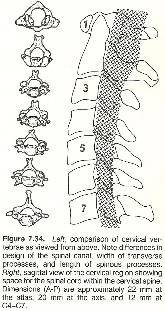
The IVD's are broader anteriorly than posteriorly to accommodate the cervical lordosis. Authorities differ as to the typical location of the nucleus
pulposus in the cervical region. Kapandji places it centrally. Cailliet places
it slightly posterior (further anterior than a lumbar nucleus), and Jeffreys
says it is distinctly posterior from the midline.
THE INTERVERTEBRAL FORAMEN
The boundaries of the cervical IVF's are designed for motion rather than
stability as compared with the dorsal and lumbar regions (Fig. 7.35). The
greatest degree of functional IVF diameter narrowing occurs ipsilaterally in
lateral bending with simultaneous extension.
THE FACET JOINTS
The articular processes incline medially in the coronal plane and obliquely
in the sagittal plane so that they are at about a 45° angle to the vertical.
Their bilateral articular surface area, which shares a good part of head weight
with the vertebral body, is about 67% of that of the vertebral body.
The short, thick, dense capsular ligaments bind the articulating processes
together, enclosing the articular cartilage and synovial tissue. Their fibers
are firmly bound to the periosteum of the superior and inferior processes and
arranged at a 90° angle to the plane of the facet. This allows maximum laxity
when the facets are in a position of rest. They normally allow no more than 2–3
mm of movement from the neutral position per segment, and possibly provide more
cervical stability than any other ligament. Capsulitis from overstretch in acute
subluxation is common. The posterior joint capsules enjoy an abundance of nociceptors and mechanoreceptors, far more than any other area of the spine.
Within the capsule, small tongues of meniscus-like tissue flaps project from
the articular surfaces into the synovial space. They are infrequently nipped" in
severe jarring at an unguarded moment during the end of extension, rotation, or
lateral bending, establishing a site of apophyseal bursitis (Fig. 7.36).
THE LOWER CERVICAL LIGAMENTS
The five lower, relatively similar, cervical vertebrae possess eight intervertebral ligamentous tissues, four posterior and four anterior. The anterior
ligaments are the anterior longitudinal ligament, the anulus fibrosus, the
posterior longitudinal ligament, and the intertransverse ligament. The posterior
ligaments are the ligamentum flavum, the capsular ligaments, and the interspinous and supraspinous ligaments.
The anterior longitudinal ligament rides close to the anterior vertebral
bodies and blends with the anulus as it crosses the IVD space. It is quite thin,
translucent, and thickest and widest over the anterior anulus. It tends to limit
extension, as does the anulus.
The posterior longitudinal ligament is firmly attached to the IVD but
separated from the vertebral bodies (except the lips) by the retrocorporeal
nutrient vessels. By not following the concavity of the vertebral bodies, the
posterior longitudinal ligament offers a smooth anterior wall for the spinal
cord. However, thickening or ossification of this ligament can encroach upon the
vertebral canal. It is much thicker than its anterior counterpart, but as
its counterpart, it is widest as the disc level. It tends to limit flexion, as
does the anulus.
The thin, fibrous, intertransverse ligament runs longitudinally between adjacent transverse processes, just anterior to the vertebral artery, joining the
anteroinferior aspect of the transverse process above to the anterosuperior lip
of the transverse process below. It serves to limit contralateral lateral bending and rotation.
The strong, thick, elastic ligamentum flavum connects the lamina of adjacent
vertebrae, riding essentially within the vertebral canal. Its usually great
elasticity prevents buckling that would impinge upon the contents of the spinal
canal (Fig. 7.37).
The interspinous ligament and the supraspinous ligament are poorly developed
in the upper cervical region. In the lower levels, the supraspinous ligament is
continuous with the ligamentum nuchae posteriorly and continuous with the interspinalis ligaments anteriorly. The supraspinous ligaments overlap and obliquely cross the midline, attaching themselves to the cervical spinous processes. The interspinous and supraspinous ligaments tend to check flexion, rotation, and anterior displacement during flexion.
The inelastic ligamentum nuchae extends in the posterior midline from the
vertebra prominens to the occiput, blending with the posterior edge of the
interspinous ligament (Fig. 7.38). It is poorly developed in humans as compared
to most other mammals, yet it serves as a cervical strap that is a mechanism of
defense against flexion injuries of the intrinsic muscles and structural displacement. When it degenerates (eg, old age), the head droops forward from the
trunk and the cervical curve straightens.
Kinematics of the Lower Cervical Spine
The IVD's contain an exceptional amount of elastin, which allows the IVD's
to conform to the many possible planes of movement. Excessive flexion is limited
by the ligamentous and muscular restraints on the separating posterior arches,
and overextension is limited by bony apposition. Other factors include the
resistance of the anular fibers to translation, the stiffness property of the
anulus relative to its vertical height, and the physical barrier produced by the
uncinate processes that are fully developed in late adolescence.
BIOMECHANIC LOWER CERVICAL INSTABILITY
Subtle instability is rarely obvious in the ambulatory patient. The most
important stabilizing agents in the mid and lower cervical spine are the anulus
fibrosus, the anterior and posterior ligaments, and the muscles, especially,
which serve as important contributing stabilizers. Upon dynamic palpation, any
segmental motion exceeding 3 mm should arouse suspicions of lack of ligament
restraint.
Neurologic Deficit. There is a rough correlation between the degree of
structural damage present and the extent of the neurologic deficit. This is more
true in the lower cervical area than that of the upper region where severe damage may appear without overt neurologic signs. In either case, however, it is
doubtful that such a deficit would exhibit without an unstable situation existing. It is not unusual for a patient to exhibit a neurologic deficit without
static displacement; ie, the vertebral segment has rebounded back into a normal
position of rest.
FACET ACTION
In the middle and lower cervical areas, A–P motion is a distinctly gliding
translation because of the 45° facet planes and the A–P biconcave discs and vertebral bodies. During flexion and extension, the superior vertebra's inferior
facets slide anterosuperior and posteroinferior on the inferior vertebra's
superior facets. During full flexion, the facets may be almost if not completely
separated. It is for this reason that an adjustment force is usually contraindicated in the fully flexed position. The center of motion is often described as
being in the superior aspect of the body of the subjacent vertebra.
Some pivotal tilting of the superior facets, backward in extension and forward in flexion, is also normal near the end of the range of motion. The facets
also tend to separate (open) on the contralateral side of rotation and lateral
bending. They approximate (jam) during extension and on the ipsilateral side of
rotation and lateral bending. Likewise, the foramina normally open on flexion,
narrow on extension, and close on the concave side of lateral flexion. Because
of the anterosuperior slant of the lower cervical facets, an inferior facet that
moves downward must also slide posterior, and vice versa.
Any corrective adjustment must take into consideration the overall degree of
the cervical lordosis, the planes of articulation, the facet tilting present,
and the degree of facet opening, as well as any underlying pathologic process
involved, and applying just enough force to overcome the resistance of the fixation.
COUPLING PATTERNS
During lateral bending, the vertebral bodies tend to rotate toward the concavity while the spinous processes swing in a greater arc towards the convexity.
Note that this is exactly opposite to the coupling action in the lumbar spine.
During cervical bending to the right, for example, the right facet of the
superior vertebra slides down the 45° plane toward the right and posterior and
the left facet slides up the 45° incline toward the left and anterior. This
coupling phenomenon is seen in circumstances in which an unusual ratio of axial
rotation and lateral bending produces a subluxation or unilateral facet dislocation.
The amount of cervical rotation that is coupled with lateral flexion varies
with the segmental level. At C2, there is 1° of rotation with every 1.5° of
lateral flexion. This 2:3 ratio changes caudally so that the degree of coupled
rotation decreases. For example, at C7, there is 1° of rotation for every 7.5°
of lateral flexion, a 2:15 ratio.
RANGE OF MOTION
All cervical vertebrae from C2 to C7 partake in flexion, extension, rotation, and lateral flexion, but some segments (eg, C5) are more active than
others. In the C3–C7 area, flexion and extension occur as slight gliding translation of the upper on the lower facets, accompanied by disc distortion. The
site of greatest movement in flexion is near the C4–C5 level (39°), while extension movement is fairly well diffused. This fact probably accounts for the high
incidence of arthritis at the midcervical area. Rotation is greatest near the
C5–C6 level (34°), slightly less above (26°–28°) and considerably less below
(13°–15°). Lateral bending in greatest near the C2–C3 level (20°) and is diminished caudally (15°–17°). The arc of lateral motion is determined by the planes of the covertebral joints (Fig. 7.40).
MOTION OF THE TRANSITIONAL CERVICOTHORACIC AREA
In the cervicothoracic area, normal movement is somewhat similar to that in
the lumbosacral area insofar as the type of stress (not magnitude of load) to
which both areas are subjected is similar. L5 is relatively immobile on the
sacrum and C7 is relatively immobile on T1, with the major amount of movement in
the cervicothoracic junction being at C6–C7 and primarily that of rotation.
REVERSAL OF THE NORMAL CERVICAL CURVE
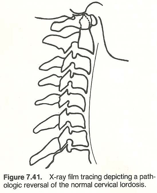
A pathologic straightening of the normal anterior curve of the cervical
spine, as viewed in a lateral weight-bearing x-ray film, results in mechanical
alteration of normal physiologic and structural integrity (Fig. 7.41). The normal vertical A–P line of gravity, as viewed laterally, falls approximately
through the odontoid and touches the anterior border of T2. As the cervical
spine tends to flatten in the erect position, the gravity line passes closer to
the center of the cervical discs.
Etiology. While the cervical curve is the first secondary curve to develop
in the infant, its maintenance in the erect posture is essentially determined by
the integrity of the lumbar curve. A flattened cervical spine that is not compensatory to a flattened lumbar spine is usually the result of a local disorder
such as a subluxation syndrome caused by posterior shifting of one or more disc
nuclei, hypertonicity of anterior musculature, or anterior ligamentous shortening as the result of local overstress, inflammation, occupational posture, or
congenital anomaly.
Symptoms and Signs. Cervical flattening is usually the result of paraspinal
spasm secondary to an underlying injury, irritation, or inflammatory process.
The acute clinical picture is one of torticollis. Other manifestations include
headaches (occipital, occipital-frontal, supraorbital), vertigo, tenderness elicited on lateral C4–C6 nerve roots, neuritis involving branches of the brachial
plexus due to nerve-root pressure, hyperesthesia of one or more fingers, and
loss or lessening of the biceps reflex on the same or contralateral side. In
rare cases, the triceps reflex may be involved. One or more symptoms are frequently aggravated by an abnormal position of the head such as during reading in
bed, an awkward sleeping position, or long-distance driving.
Roentgenographic Considerations. Rehberger reports the typical radiographic
findings to include loss of the normal lordotic curve by the straightened cervical spine (78% cases), anterior and posterior subluxation on flexion and
extension views, narrowing of IVD spaces at C4–C6 in 46% cases, discopathy at
the affected vertebral level as the injury progresses, and osteoarthritic
changes which are often accompanied by foraminal spurring.
Biomechanics. A flattened cervical spine in the erect posture resembles a
normal spine during flexion. To appreciate the mechanisms involved, it is well
here to review the biomechanics involved. The nucleus of the disc serves as a
fulcrum during flexion and return extension. When the spine is subjected to
bending loads during flexion, half of the disc on the convex side suffers tension, widens, and contracts, while the other half of the disc on the concave
side suffers compression, thins, and bulges. Concurrently, the nucleus bulges on
the side of tension and contracts on the side of compression, which increases
tension on the adjacent anulus. This creates a self-stabilizing counteracting
flexion force to the motion unit that aids a return to the resting position.
Case Management. Specific correction of offending vertebral subluxations
should be accomplished. Adjunctive care includes massage and methods to reduce
muscle spasm such as ultrasound, diathermy, hydrocollator packs, reflex spinal
techniques, and a rolled towel placed under the neck in the supine position to
increase the cervical curve. The individual should be instructed to sleep without a pillow. Cervical muscle re-education is quite helpful.
Prognosis. Rehberger and Barge report that the prognosis is excellent if the
condition is treated early and the case is not complicated by fracture or dislocation, but guarded if the trauma is severe. In cases of minimal cervical
discopathy, at least symptomatic relief can be expected. Prognosis is poor in
advanced degenerative osteoarthritis.
Lower Cervical Trauma
Cervical fractures and dislocations are not common except in the elderly
where a degree of osteoporosis is evident. They are usually the result of severe
trauma. Bruises on the face, scalp, and shoulders may offer clues as to the
mechanism of injury. Signs of vertebral tenderness, limitation in movement,
muscle spasm, and neurologic deficit should be sought. As in upper-cervical
damage, careful emergency management is necessary to avoid paralysis and death.
Fracture and/or dislocation of any cervical vertebra require hospitalization for
reduction, bone traction, and casting. Keep in mind that overdiagnosing instability of C2–C3 is a common pitfall.
SUBLUXATIONS
Due to the planes of normal articular processes, a straight horizontal
subluxation is an anatomic impossibility unless there is a fracture of the articular processes. The body of any lower cervical vertebra follows the planes of
the covertebral and posterior facets in movement. If a spinous process moves
left, it does so by inscribing an arc toward the superior and anterior while
simultaneously the right transverse process moves inferiorly and somewhat
posteriorly. It is thus impossible for an individual vertebra to be rotated
straight right or left on its longitudinal axis, and irrational to make a
listing of right or left. A vertebra cannot be subluxated without one of the
articular processes moving either superiorly or inferiorly; thus it can be said
that superiority or inferiority attends every posterior/anterior subluxation.
It should be kept in mind that the nerve root is anterior and inferior to
the facets in the cervical spine. If subluxation of a vertebra occurs in a
superior direction, the contents of the IVF become stretched because elongating
and narrowing the vertical diameter of the IVF will cause traction upon the
nerve trunk plus compression against the anterior portion of the foramen. If
there is subluxation in an inferior direction, shortening and widening of the
foramen occurs. Because the nerve sheath is often firmly anchored by tissues
connecting it to the borders of the foramen in the adult, a stretching effect is
exerted on the nerve sheath whenever its shape is altered. It can thus be appreciated that enlarging the IVF can cause as much trouble as a reduction in the
size of the IVF. Also, it is impossible to subluxate a vertebra between C2 and
L5, inclusive, without changing the shape of its IVD in compensation.
A subluxation of one or more of the lower cervical vertebrae often involves
the brachial plexus.
Table 7.5 lists the nerves of the plexus and their function.
Inasmuch as the distribution of the brachial plexus is so extensive, a multitude of abnormal reflections may be seen in its areas of distribution which
must be appreciated by knowledge of the pathophysiology involved. A few of the
more common disturbances caused by lower cervical subluxations would include
shoulder neuralgias, neuralgias along the medial arm and forearm or elbow, unclassified wrist drop and hand dystrophies, acroparesthesia, weak grip strength,
and vague "rheumatic" wrist or hand complaints. A subluxation of one or more of
the C3, C4, or C5 segments may involve the phrenic nerve and produce symptoms of
severe chronic hiccup and other diaphragmatic disorders.
GENERAL ASPECTS OF FRACTURES AND DISLOCATIONS
Isolated fractures following trauma occur at all levels of the cervical
spine. Vertebral body fractures, however, occur most frequently at C6 and C7 and
least frequently at C4. The four common types of vertebral body fractures are
anterior marginal fractures from A–P forces, comminuted fractures from axial
forces, and lateral wedge fractures and uncinate process fractures from lateral
stress. Vertical compression or flexion compression damage (Fig. 7.42) is sometimes seen, but extension injuries (eg, whiplash) are more common. Spinous process fractures usually occur at the C6 or C7 level after acute flexion or a blow
to the flexed neck producing ligamentous avulsion. There is immediate "hot" pain
in the area of the spinous process which is increased by flexion. Any injury to
C6–C7 is difficult to view on film because of overlapping structures.
It is most difficult to conceive of a vertebral body compression fracture
not being secondary to severe end-plate fracture, even if roentgenographic evidence of end-plate failure is not seen. Invariably, the end-plate must fail
first in a healthy vertebra subjected to extreme vertical and/or bending forces.
Flexion Injuries. In a blow to the occiput directed upward, the posterior
elements receive the greatest trauma because of the shear component in the
hyperflexion force. During forceful cervical flexion, a unilateral facet dislocation and/or fracture may occur with the contralateral side remaining intact,
especially if the force is oblique. Bilateral dislocation or fracture-dislocation may occur if the facets are forced to override without rotation (Fig.
7.44). Unilateral dislocation is more common in the lower cervical area than in
the upper area.
Extension Injuries. Forceful extension can produce tearing of the anterior
longitudinal ligament and anterior anulus which may coexist with an avulsion
fracture at the lips of the anterior vertebral body (Fig. 7.45). If rupture
occurs, further force is absorbed by the articular processes, spinous processes,
laminae, and pedicles, in that order. About 50% of all cervical fractures are
of the vertebral arch. If the articular processes fracture and the posterior
arch fails, the vertebral body will inevitably be displaced anteriorly. Transverse pedicle fracture or severe posterior subluxation may also occur. Keep in
mind that the articular pillars of the C3–C7 vertebrae are not designed as the
lateral masses of the atlas. These pillars project laterally on each side at the
junction of the lamina and pedicle.
Tenderness will usually be shown along the lateral musculature. Upper extremity pain or numbness and restricted cervical motion at one or more interspace
during flexion-extension may be exhibited. Neurologic symptoms may be severe and
prolonged without demonstrable roentgenographic evidence. Cord damage without
apparent structural damage may result from a bulge created by a buckled degenerated (nonelastic) ligamentum flavum at the posterior of the spinal canal (Fig.
7.46). The cord may also be pinched between the posteroinferior edge of the
superior vertebral body and the laminae of the inferior segment.
Lateral Flexion Injuries. When the head is forced to severely tilt laterally, there is always a coupled component of rotation involved. Compression
wedging of structures on the concave side occurs, and tension on the structures
on the convex side is produced. There are four typical severe traumatic effects
throughout the cervical area:
(2) a unilateral compression fracture of the vertebral body will occur;
(3) there will be fracture of the uncinate or transverse process or fracture and/or
dislocation of the articular process ipsilaterally with ligamentous rupture
contralaterally;
(4) there will be brachial plexus avulsion, possibly associated
with a cervical and/or thoracic fracture.
(2) the posterior ligaments, posterior joint capsules, and posterior longitudinal ligament (in that order) rupture when the neck is overflexed and
rotated.Priority Priority
Suspect Suspect
Nerve(s) Area of Localized Pain Nerve(s) Area of Localized Pain
Trigeminal Anterior head and face T5–12 Peritoneum
C1–2, T7–12 Occiput T6–10 Pancreas, spleen
C2–3 Forehead T7–9 Ascending colon
C3, T1–5 Neck T8–9 Gallbladder
C3–4, T1–3 Aortic arch T9–10 Small intestines
C3–4, T1–5 Heart T9–11 Transverse colon
C3–4, T1–8 Head and face T10–11 Umbilical area, ovary,
C3–4, T3–5 Lungs testicle
C3–4, T6–7 Stomach, cardiac aspect T10–12 Crown of head, scrotum,
C3–4, T8–10 Stomach, pyloric aspect lower limbs
C3–4, T7–9 Liver T10–12, S1–3 Prostate
C4 Shoulder girdle, temple T10–L1 Kidney, uterine body
area T11–L1 Urethra, epididymis
C5 Deltoid area T11–L2 Bladder neck, descend-
C6 Thumb ing colon
C7 First or index finger T11–L1 Suprarenal area
C8 Fourth finger T12–L1, S1–4 Uterine neck
T1 Fifth finger T12–L2 Ureter
T1–4 Thorax L1 Groin
T2 Nipple area L1–3, S1–4 Bladder body, rectum,
T2–4 Bronchi genital organs
T2–5 Upper limbs L3 Knee, medial aspect
T2–12 Pleura L5 Great toe
T4–5 Mammae bodies S1 Fifth toe
T4–7 Thoracic aorta S2 Thigh, posterior aspect
T5–8 Esophagus (caudal) S2–4 Cervix
Note: Authorities differ somewhat as to exact levels, and variances of a segment
above or below are commonly stated by different authorities. The above data are
a composite of the findings from several sources (Courtesy of Associated Chiropractic Academic Press).
Cervical Subluxation Syndromes
Subluxations, regardless of region, are difficult to classify under normal
categories of trauma because they can involve bone, joint, muscle, ligament,
disc, nerve, cord, lymphatic and vascular tissues. Thus, subluxation is a finding and a syndrome and not a diagnosis.
Once a vertebra loses its ideal relationship with contiguous structures and
becomes relatively fixed at some point within its normal scope of movement, it
is no longer competent to fully participate in ideal coordinated spinal movement. The affected area becomes the target for unusual weight bearing and traumatic stress. In addition to attending circulatory and static changes in the
involved area, there is disturbed neural activity that may be exhibited as
changes in superficial and deep reflexes, tremors and spasms, hyperkinesia,
pupillary changes, and excessive lacrimation.
PERTINENT FUNCTIONAL ANATOMY OF THE CERVICAL PLEXUS
The dura mater of the spinal cord is firmly fixed to the margin of the foramen magnum and to the 2nd and 3rd cervical vertebrae. In other spinal areas, it
is separated from the vertebral canal by the epidural space. Since both the C1
nerve and the vertebral artery pass through this membrane and both are beneath
the superior articulation of the atlas and under the overhanging occiput,
atlanto-occipital distortion may cause traction of the dura mater producing
irritation of the artery and nerve unilaterally and compressional occlusion contralaterally. De Rusha feels that this helps us understand those cases of suboccipital neuralgia where a patient upon turning his head to one side increases
the headache and vertigo that are relieved when the head is turned to the opposite side.
There is also a synapse between the upper cervical nerves and the trigeminal
nerve, which also supplies the dura mater. This may explain why irritation of C1
results in a neuralgia not only confined to the base of the skull but is also
referred to the forehead or eye via the supraorbital branch of the trigeminal.
The greater occipital (C2) nerve does not tend to do this. It exits between the
posterior arch of the atlas and above the lamina of the axis (Fig. 7.48), referring pain to the atlanto-occipital area (Fig. 7.30) and often to the vertex of
the head.
The superficial sensory cutaneous set of the cervical plexus (C1–C4) is
frequently involved in subluxations of the upper four segments (refer to Table
7.4), particularly when there are predisposing spondylitic degenerative changes.
Janse describes four resultant neuralgias:
(2) greater auricular nerve neuralgia, extending in front and behind the auricle, skin over the parotid gland,
paralleling the distribution of the auriculotemporal branch of the trigeminus
and easily misdiagnosed as chronic trifacial neuralgia;
(3) cervical cutaneous
nerve neuralgia, involving the area of the middle third of the platysma to the
midline, possibly extending from the chin to the sternum;
(4) supraclavicular
nerve neuralgia, depending upon which rami are affected, the neuralgia may
involve the suprasternal area, pectoral area, or deltoid area. Thus, sternoclavicular and acromioclavicular neuralgias may originate in the spinal levels
of the supraclavicular nerve.
Irritative lesions involving the cervical articulations may in turn irritate
the sympathetic nerve plexuses ascending into the head via the vertebral and
carotid arteries. Some cases of visual and aural symptoms are related to upper
cervical distortion where the arch of the atlas snugly hugs the occiput, thus
possibly irritating the sympathetic plexus near the vertebral artery as well as
partially compressing the vessel. To appreciate this, note that the visual cortical area of the occipital lobe requires an ideal blood supply dependent on the
sympathetics ascending the great vessels of the neck, and this holds true for
the inner ear as well. To test this syndrome, De Rusha suggests having the
supine patient read some printed matter while the examiner places gentle traction on the skull, separating the atlanto-occipital articulations. A positive
sign is when the patient, often to his surprise, experiences momentarily enhanced visual acuity or a reduced tinnitus.
CERVICAL NERVE ROOT INSULTS
Disturbances of nerve function associated with subluxation syndromes basically manifest as abnormalities in sensory interpretations and/or motor
activities (Fig. 7.49). These disturbances may be through one of two primary
mechanisms: direct nerve or nerve root disorders, or of a reflex nature.
Motor Changes. Nerve root insults from subluxations may also be evident as
disturbances in motor reflexes and/or muscular strength. Examples of these
reflexes include the deep tendon reflexes such as seen in the reduced biceps
reflex when involvement occurs between C5–C6; or the reduced triceps reflex when
involvement occurs between C6–C7. These reflexes must also be compared bilaterally to judge whether hyporeflexia is unilateral. Unilateral hyperreflexia is
pathognomonic of an upper motor neuron lesion. Prolonged and/or severe nerve
root irritation may also cause evidence of trophic changes in the tissues
supplied.
Underlying Factors. The common subluxation picture is rarely pure. It is
often superimposed upon subclinical processes in the mature patient such as
vertebral instability, osteochondrophytic ridges at the uncovertebral joints,
apophyseal thickening and exostosis, or canal encroachment by a buckling ligamentum flavum, spinal stenosis, posterior vertebral body spurs, disc protrusions, dura and dentate thickening, arachnoid cysts, dura and arachnoid adhesions, and ossification of the posterior longitudinal ligament.
SUBLUXATION–INDUCED REFLEX SYNDROMES
Certain spinosomatic and spinovisceral syndromes may result from cervical
subluxation. For example, if the involvement is in the area of C1–C4, the cervical portion of the sympathetic gangliated chain or the 9th–12th cranial nerves
as they exit from the base of the skull and pass into their compartments within
the deep cervical fascia may be involved. The syndrome may include:
(2) greater occipital nerve extension neuralgia;
(3) mandibular, cervical, auricular, pectoral, or precordial neuralgia;
(4) paroxysmal torticollis;
(5) congestion of the upper respiratory mucosa, paranasal sinuses, or eustachian tube with hearing loss;
(6) cardiorespiratory attacks;
(7) ocular muscle malfunction;
(8) pathologic hiccups;
(9) scalenus anticus syndrome; or
(10) painful spasms in the suboccipital area.
NEUROVASCULAR IMPLICATIONS OF UPPER CERVICAL SUBLUXATIONS
Loss of mobility of any one or more segments of the spine correspondingly
influences circulation. The resulting partial anoxia has a harmful influence
upon nerve function. The artery and vein supplying a spinal nerve are situated
in the foramen between the nerve and the fibrous tissue in the anterior portion
of the foramen. It is unlikely that circulation to the nerve would be disrupted
without first irritating or compressing the nerve because the arteries and veins
are much smaller, the blood pressure within the lumen makes them resistant to
compression, and nerve tissue is much more responsive to encroachment irritation.
The Medulla Oblongata. The medulla oblongata extends well into the lower
reaches of the foramen magnum and the ligamentous ring that connects it with the
atlas, thus any type of occipital or atlantal subluxation may produce abnormal
pressure on this portion of the brain stem. Bilateral posterior shifting of the
occiput or atlas may cause pressure upon the pyramids or adjacent olivary bodies
producing a syndrome of upper motor neuron involvement characterized by a degree
of spastic paralysis or ataxia. A lateral shifting of the occiput may cause
pressure upon the tubercle of Rolando producing pain in the area of trigeminal
nerve distribution, headache, sinus discomforts, ocular neuralgias, and aches in
the jaw.
The Vertebral Arteries. Janse relates that any cervical subluxation (particularly atlantal, axial, or occipital) producing muscle spasm may produce unilateral or bilateral constriction of the vertebral arteries resulting in circulatory impairment. A large number of equilibrium, cardiac, respiratory, cranial nerve, extrapyramidal, vagal, visual, and auditory symptoms may follow.
The vertebral nerve (sympathetic) runs along the vertebral artery within the
arterial foramen of the cervical transverse processes. Irritation to this nerve
is considered to occur from mechanical irritation to the vertebral artery anywhere along its course producing symptoms of a vasomotor nature; eg, headache,
vertigo, tinnitus, nasal disturbances, facial pain, facial flushing, and
pharyngeal paresthesias. Cailliet points out that although sympathetic fibers
have not been found along the cervical roots, surgical decompression of an
entrapped nerve root relieves symptoms attributed to the sympathetics. The mechanism for this effect is unclear.
The Vertebral Veins and Deep Cervical Veins. Spasm of the suboccipital muscles may cause a decided impediment of venous drainage from the suboccipital
area via vertebral and deep cervical veins, resulting in a passive congestion
with consequent pressure upon the sensory nerve endings in the area. This is
perceived by the patient as unilateral or bilateral pain and a throbbing discomfort, and may be palpated as knotty lumps within suboccipital muscles. The condition appears to be of a reflex nature more common among people under mental
tension or those who work closely with their eyes over long periods.
Cerebrospinal Circulation. Any event that would cause constriction in the
connecting area between the cerebral subarachnoid space and the vertebral canal
limits the escape of cerebrospinal fluid into the inferior vertebral canal. This
results in a degree of increased intracranial pressure. An atlanto-occipital
subluxation may cause the dura mater of the cisterna cerebellaris to be pressed
against the posterior medullary velum and partially occlude the foramina of
Luschka and Magendie and interfere with the flow from the 4th ventricle. The
resulting increase of intraventricular fluid accumulation may create a large
variety of symptoms such as deep-seated, stubborn, "internal pressure" headaches, nausea, a tendency toward projectile vomiting, bizzare and unusual visual
disturbances, and protopathic ataxias.
Headaches of Cervical Origin
It has been the experience of Markovich, the renowned neurologist, that the
most common headache is the type caused by neuromuscular skeletal imbalance. He
points out that "the head in the human species has changed its position from the
quadruped to the erect, thereby changing the basic relationship between the cervical spine and the head, with its important functional structures, and the rest
of the body." For reference, he calls attention to the quite delicate interaction and highly sensitive biofeedback or servo-mechanisms that continually make
adjustments in body balance, vision, pressure, and hearing with head and neck
posture. "These regulatory, homeostatic mechanisms can be disturbed by a variety
of conditions, originating at any level, including the inflammation and/or irritation of the cephalic projection of the upper cervical nerves (cervico-occipital neuralgias)."
Abnormal contraction of the muscles at the occipitocervical area appears to
generate a type of "ischemic irritation" that includes the entrapment of the C2
nerves (greater and lesser occipital) as they pierce the thick muscle and ascend
to the back of the head to supply the back of the scalp, the temples, and the
ear lobes, sending branches to the top of the head, the back of the eye, and the
angle of the jaw. Differentiation of various types of headaches is shown in
Table 7.7, adapted from Markovich's data. However, keep in mind that a patient
may not exhibit such a clear-cut picture. For example, vascular migraine may be
superimposed on occipitocervical neuralgia or episodes may be interposed,
depending upon the causes involved.
Table 7.7. Differentiation of Common Types of Headaches
               Occipito-                                        Temporo-
               cervical         Trigeminal       Vascular       mandibular
Symptom/Sign   Neuralgia        Neuralgia        Migraine       Traction
Pain           Throbbing,       Excruciating,    Severe,        Severe,
               paroxysmal       paroxysmal       paroxysmal     dull

Quality        Muscle spasms    Stabbing         Throbbing       Dull ache

Location       Occipital        Facial           Unilateral      Facial

Aura           None             None             Visual          None

Duration       Days             Brief            Hours           Chronic

Associated     Earache          Trigger zones    Vomiting        Bruxism
symptoms       Eye pain                          Photophobia     Malocclusion
and signs      Neck pain                         Irritability    Earache
               Paresthesias                                      Joint clicks
               Anxiety
               Tinnitus
               Nausea
Neurovascular Compression Syndromes
The cervicothoracic junction area is a unique spinal area that receives far
less attention than it deserves in both medicine and chiropractic. It is a common site of developmental anomalies; it is a major site of arterial, lymphatic,
and neurologic traffic; and it presents the juncture of the highly mobile cervical spine with the very limited thoracic spine. This latter point is biomechanically significant.
BACKGROUND
There are several syndromes to consider under the classification of neurovascular compression syndromes (also termed thoracic outlet or inlet syndromes),
each of which may produce the symptom complex of radiating pain over the
shoulders and down the arms, atrophic disturbance, paresthesias, and vasomotor
disturbances. These syndromes, however, do not necessarily indicate the specific
cause of the problem.
Incidence. The 4th and 5th decades mark the highest incidence of trouble in
this area, probably because of regressive muscular changes. The incidence of
disorders is more frequent in females in the order of 3:1. This is probably due
to heavier upper-extremity work.
Etiology. Trauma to the head, neck, or shoulder girdle is a common factor.
In some cases, poor posture, anomalies, or muscle contractures may be involved.
Reduced tone in the muscles of the shoulder girdle, by itself, has been shown to
allow depression of the clavicle that narrows the thoracic outlet and compresses
the neurovascular bundle. In addition, subluxation syndromes (eg, retrolisthesis) may initiate these and other disturbances of the shoulder girdle and must
be further evaluated. Cervical pathology such as spinal canal or IVF encroachment by a buckling ligamentum flavum, spinal stenosis, or spurs should be a consideration. During degeneration, the dura and dentates become thickened, dura
and arachnoid adhesions become prevalent, osteochondrophytes may develop from
the borders of the canal or foramen –all of which tend to restrict the cord
and/or nerve root during cervical motions. Osteochondrophytes near the foramen
can readily compress the vertebral artery and root together.
Grieve calls attention to "the poorly recognized part played by minor unilateral joint abnormalities of the upper three or four ribs, possibly often by
tension of soft tissue attachments as a consequence of cervical joint irritability at higher segments, or due to the chronic sequelae of trauma...."
SYMPTOMS AND SIGNS
Symptoms usually do not occur until after the ribs have ossified. Two groups
of symptoms are seen, those of scalenus anticus syndrome and those due to cervical rib pressure. The symptoms of cervical rib and scalenus syndrome are similar, but the scalenus anticus muscle is the primary factor in the production of
neurocirculatory compression whether a cervical rib is present or not.
When symptoms are present, they are usually from compression of the lower
cord of the brachial plexus and subclavian vessels such as numbness and pain in
the ulnar nerve distribution. Pain is worse at night because of pressure from
the recumbent position, and its intensity varies throughout the day. Tiredness
and weakness of the arm, finger cramps, numbness, tingling, coldness of the
hand, areas of hyperesthesia, muscle degeneration in the hand, a lump at the
base of the neck, tremor, and discoloration of the fingers are characteristic.
Work and exercise accentuate symptoms, while rest and elevation of the extremity
relieve symptoms. Adson's and other similar compression signs will usually be
positive.
Anatomic Considerations. Working above and behind to produce shoulder abduction and retraction (eg, painting a ceiling, repairing a ceiling fixture) will
definitely cause temporary clavicle encroachment on the brachial plexus and
press the subclavian artery against the scalenus medius. For many years, the
cause was attributed to costoclavicular compression from shoulder depression.
However, postmortum stress tests have shown the following:
Clinical Compression Tests
X-ray films should always be taken before performing a cervical compression
test, especially when the patient has experienced trauma or shows physical signs
of advanced degeneration. It is important to rule out possible conditions that
would be aggravated by any testing procedure.
ACTIVE CERVICAL ROTARY COMPRESSION TEST
With the patient seated, he should be observed while voluntarily laterally
flexing his head toward the side being examined. With the neck flexed, the
patient is then instructed to rotate his chin toward the same side, which narrows the IVF diameters on the side of concavity. Pain or reduplication of other
symptoms probably indicates a physiologic narrowing of one or more IVF's.
PASSIVE CERVICAL COMPRESSION TESTS
Two tests are involved. First, with the patient seated, the examiner stands
behind the patient. The patient's head is laterally flexed and rotated slightly
toward the side being examined. Interlocked fingers are placed on the patient's
scalp and gently pressed caudally. If an IVF is physiologically narrowed, this
maneuver will further insult the foramen by compressing the disc and narrowing
the foramen, causing pain and reduplication of other symptoms. In the second
test, the patient's neck is extended by the examiner who then places interlocked
hands on the patient's scalp and gently presses caudally. If an IVF is physiologically narrowed, this maneuver mechanically compromises the foraminal diameters
bilaterally and causes pain and reduplication of other symptoms.
SPURLING'S TEST
This is a variation of the passive cervical compression test. The patient's
head is turned to the maximum toward one side and then laterally flexed to the
maximum. A fist is placed on the patient's scalp, and a moderate blow is delivered to it by the other fist. The patient's position produces reduced IVF spaces, and the blow causes a herniated disc to bulge further into the IVF space or
an irritated nerve root to be aggravated, thus increasing the symptoms.
SHOULDER DEPRESSION TEST
With the patient seated, the examiner stands behind the subject. The
patient's head is laterally flexed away from the side being examined. The doctor
stabilizes the patient's shoulder with one hand and applies pressure alongside
the patient's head with the palm of the other hand; stretching the dural root
sleeves and nerve roots or aggravating radicular pain if the nerve roots adhere
to the foramina. Extravasations, edema, encroachments, and conversion of fibrinogen into fibrin may result in interfascicular, foraminal, and articular adhesions and inflammations that will restrict fascicular glide and the ingress and
egress of the foraminal contents. Thus, pain and reduplication of other symptoms
during the test suggest adhesions between the nerve's dura sleeve and other
structures around the IVF.
CERVICAL DISTRACTION TEST
With the patient seated, the examiner stands to the side of the patient and
places one hand under the patient's chin and the other hand under the base of
the occiput (Fig. 7.50). Slowly and gradually the patient's head is lifted to
remove weight from the cervical spine. This maneuver elongates the IVF's, decreases the pressure on the joint capsules around the facet joints, and
stretches the paravertebral musculature. If the maneuver decreases pain and
relieves other symptoms, it is a probable indication of narrowing of one or more
IVF's, cervical facet syndrome, or spastic paravertebral muscles.
ADSON'S TEST
With the patient seated, the examiner palpates the radial pulse and advises
the patient to bend his head obliquely backward to the opposite side being examined, take a deep breath, and tighten the neck and chest muscles on the side
tested. The maneuver decreases the interscalene space (anterior and middle scalene muscles) and increases any existing compression of the subclavian artery
and lower components (C8 and T1) of the brachial plexus against the 1st rib.
Marked weakening of the pulse or increased paresthesiae indicate a positive sign
of pressure on the neurovascular bundle, particularly of the subclavian artery
as it passes between or through the scaleni musculature, thus indicating a probable cervical rib or scalenus anticus syndrome.
EDEN'S TEST
With the patient seated, the examiner palpates the radial pulse and instructs the patient to pull the shoulders backward, throw the chest out in a
"military posture," and hold a deep inspiration as the pulse is examined. The
test is positive if weakening or loss of the pulse occurs, indicating pressure
on the neurovascular bundle as it passes between the clavicle and the 1st rib,
and thus a costoclavicular syndrome.
WRIGHT'S TEST
With the patient seated, the radial pulse is palpated from the posterior in
the downward position and as the arm is passively moved through an 180° arc. If
the pulse diminishes or disappears in this arc or if neurologic symptoms develop, it may indicate pressure on the axillary artery and vein under the pectoralis minor tendon and coracoid process or compression in the retroclavicular
spaces between the clavicle and 1st rib, and thus be a hyperabduction syndrome.
Vertebrobasilar System Patency Tests
Although cerebrovascular accidents are extremely rare following cervical
manipulation, a few cases have been reported that justify careful evaluation
prior to cervical manipulation. Bergmann describes four clinical tests to evaluate the patency of the vertebrobasilar system.
MAIGNE'S TEST
The examiner places a seated patient's head in extension and rotation. This
position is held for about 15–40 seconds on each side. A positive sign is indicated by nystagmus or symptoms of vertebrobasilar ischemia.
NOTE:
Maigne's, and the following test, puts the Vertebral Artery in the most compromised position possible. BEFORE using them, please review Chiro.Org’s Stroke Page, in particular the section on the 5 D's and the 3 N's testing. To the extent that is possible, these tests may reveal an occult TIA or stroke, while in its prodromal state, so that you can refer that patient for vascular assessment.
DeKLEYN'S TEST
The patient is placed supine on an adjusting table, and the head rest is
lowered. The examiner extends and rotates the patient's head, and this position
is held for about 15–40 seconds on each side. A positive sign is the same as
that in Maigne's test.
HAUTANT'S TEST
The examiner places a seated patient's upper limbs so that they are abducted
forward with the palms turned upward (supination). The patient is instructed to
close his eyes, and the examiner extends and rotates the patient's head. This
position is held for about 15–40 seconds on each side. A positive sign is for
one or both arms to drop into a pronated position.
UNDERBURGER'S TEST
The patient is asked to stand with his upper limbs outstretched, his eyes
closed, and then to march in place with his head extended and rotated. The examiner should stand close to the patient during the test because a positive sign
is a loss of balance.
Visual Subluxation Patterns
Clinical postural patterns reflect an individual's biomechanics that are
responsive to underlying physiologic processes. This is readily appreciated clinically in many acute conditions such as the antalgic positions of sciatica,
extremity dislocation, the opisthotonus of spinal meningitis, the orthopnea of
asthma, the emprosthotonus of tetanus, the pleurothotonus of strychnine poisoning, the rigid abdominal spasm of peritonitis, and other postures that a patient
might assume to gain relief. However, the postures reflected in chronic subluxations are much more subtle and often assumed unconsciously via proprioceptive
mechanisms.
The analysis of "body language" opens the way for much subjective interpretation, and there is a tendency to point to signs that confirm one's interpretation and ignore those that do not. Nevertheless, as shown in our previous
discussion of gait, many specific neuromusculoskeletal disorders can be accurately judged by observing the body in stance and in action. The interpretation of
spinal distortion patterns is likewise an art, and, as in any diagnostic art,
its effectiveness is in knowing the normal from the abnormal and knowing where
the normal or abnormal begins and ends.
Several chiropractic researchers feel that many postural distortions are
essentially massive movement of structure held in fixation by impulse bombardment initiated by neural excitement. In this sense, a visual pattern is a clue
that must be confirmed by other diagnostic methods such as static and dynamic
palpation and roentgenography. Other researchers feel that it is most difficult
to relate specific subluxations to gross postural patterns.
In this section and especially in the future chapters on the thoracic spine
and the lumbar spine and pelvis, we will offer some of the basic premises of
visual subluxation patterns with the hope that it will stimulate further research in spinal analysis.
CERVICAL SUBLUXATION PATTERNS
In normal neutral posture, the ears (semicircular canals) and eyes will be
held level and parallel to the shoulders for resting orientation. Inspection
is best made by using a double plumb line or a transparent grid and a foot-stabilizing device. De Jarnette feels that practically all subluxation patterns
have some effect upon the occiput and upper cervical spine. Any shifting of the
cranial mass (eg, tilt) will have an affect upon the cervical muscles which, in
turn, alter the stabilization of the position of the head upon the cervical
spine. When the occiput, atlas, and axis are in good relationship, the ears will
appear level and equally prominent, reflecting equal weight distribution bilaterally and good functioning of the righting mechanism (Fig. 7.51).
OCCIPITAL SUBLUXATION PATTERNS
When viewed from the posterior, an occiput inferior on the right due to a
compressed condyle, for example, will present a head tilt low on the right. The
ears will be equally prominent. A purely rotational subluxation of the occiput
on the atlas will exhibit a level skull with the ear on the side of relative
posteriority more prominent (Fig. 7.52).
ATLAS SUBLUXATION PATTERNS
It is theorized by many that every atlas subluxation changes the way that
the skull adapts to body posture. When there is an occipital tilt and the ear is
more prominent (rotated posteriorly) on the side of occipital superiority, an
ipsilateral posterior or contralateral anterior atlas subluxation is suspect
(Fig. 7.53).
When the atlas is fixed in a posterior rotation on the right or an anterior
rotation on the left, the right ear and mandible will be more prominent (rotated
posteriorly) than the left when viewed from the posterior. The prominence of the
mandible is said to be due to the subluxation's effect upon the axis.
An atlas fixed in abnormal lateral flexion will present a tipped occiput
shown by unlevel ears, but the ears will be equally prominent. This is the same
picture of that of a contralateral sunken condyle. With such a subluxation,
reports De Jarnette, the shoulders will be level or one will be slightly low on
the side of atlas inferiority if shoulder problems are associated. That is, the
arm will be carried low on the side of atlas side slip.
AXIS SUBLUXATION PATTERNS
Rotary subluxations of the axis also appear to present a typical picture. An
axis subluxation changes the way the skull balances upon the atlas. If the axis
is fixed posterior and inferior on the right, for example, the right ear will be
rotated posteriorly and be more prominent, and the skull will be tipped inferior
on the right when viewed from behind (Fig. 7.54). A unilaterally prominent mandible is thought to be an effect only in axis subluxations or when the axis
influences an atlas subluxation.
CERVICAL SUBLUXATIONS WITH SCIATICA SYNDROMES
It is not unusual to see cervical complaints associated with a case of true
sciatica. In right sciatica, for example, the patient's stance may exhibit very
little distortion when viewed from the posterior. If the patient is asked to
raise his left heel, however, a pronounced inferior tilt of the skull on the
left may appear with the dorsolumbar spine forming a mild left scoliosis. When
the patient is asked to lower his left heel and raise his right heel, the dorsolumbar curve increases, an acute right dorsocervical scoliosis develops, and
the occipital tilt is increased (Fig. 7.55). The raising of the right heel
invariably increases right sciatic pain, according to De Jarnette, and also produces cervical pain. This manifestation is said to indicate a primary or secondary cervical subluxation, but other means will be necessary to isolate the
exact segmental level of involvement.
Postural Realignment
Because the center of gravity of the head lies anterior to the occipital
condyles, definite force must be applied by the posterior muscles of the neck to
hold the head erect. In addition, several groups of muscles are attached
directly or indirectly to the anterior part of the head, and their function adds
to the force of gravity to increase the load on the posterior cervical musculature. Gelb feels that the most important anterior neck muscles are the masticatory and supra- and infra-hyoid groups, which constitute a sort of chain with
the hyoid bone and mandible to which they are attached. They join the head to
the shoulder girdle anteriorly.
Deformity of cervical posture can be associated with or without neck pain.
As discussed, the deformity may depict curvature exaggeration, flattening, occipital tilt, rotation, and areas of complete or partial segmental fixation. If
pain is present, deviation may be toward or away from the site of pain, depending upon the primary site of irritation. Keep in mind that spinal stability is
essentially under the control of neuromuscular mechanisms rather than the ligaments.
Although segmental spasm or instability may not be detectable by observation, it usually becomes evident by alert dynamic palpation. Careful localization of motion and muscle strength is necessary prior to prescribing any exercise program to assist postural realignment.
PROPRIOCEPTION
In general, the abundant proprioceptors of the vertebral column enable the
brain to know where each segment is and what it is doing at any given time
without visual confirmation. More specifically, data are relayed as to the
degree of muscle tension and/or the length of muscles via the muscle spindles
and Golgi tendon organs. Tension messages are moved through fast-conducting nerves from annulospiral endings and through higher threshold nerves from flower
spray receptors in the muscle spindle. The less complex Golgi tendon organs near
the musculotendinous junctions discharge impulses initiated by either muscle
contraction or stretch. Other receptors near the articular surfaces relate
messages about joint speed and direction of motion.
In postural realignment, as Gelb points out, restoring muscles to their physiologic resting length is a three-dimensional concept, requiring placing the
origin and insertion of muscles in a correct three-dimensional relationship.
As mentioned previously, Lieb shows a number of full spine x-ray prints of
severe spinal distortions greatly improved through improvement of dental occlusion. If the temporomandibular joint has such a proprioceptive influence upon
posture, it is no wonder that chiropractors who specialize in solely upper-
cervical or sacral correction exhibit a likewise abundance of such before-and-
after exhibits inasmuch as each spinal segment is richly endowed with equal or
greater receptors than that of the jaw.
THE CERVICAL SPINAL RECEPTORS
The apophyseal joints of the cervical spine are richly innervated with
mechanoreceptors and afferent fibers, endowed more than any other spinal region.
Activity from the cervical articular receptors exerts significant facilitatory
and inhibitory reflex effects on the muscles of the neck and both the upper and
lower extremities.
Wyke points out that the patterns of "normal cervical articular mechanoreceptor reflexes are profoundly distorted when cervical articular nociceptive
afferent activity is added to that derived from the normally functioning cervical mechanoreceptors." To underscore this point, he states that:
(2) arm movement control in the absence of visual aid is considerably affected by rotation of the head,
and
(3) induced unilateral local anesthesia of the cervical joints in healthy
subjects produces severe postural instability, dizziness, nystagmus, and muscular incoordination. These signs and symptoms are similar to those experienced by
some patients who suffer from cervical spondylosis, ankylosing spondylitis,
gross fixations, and some while wearing an orthopedic cervical collar.
RIGHTING MECHANISMS
Several head extensors arise from the lower cervical and upper thoracic vertebrae that exert an oblique posteroinferior pull on the occiput. If the line of
pull falls behind the atlanto-occipital joint, a rotary movement results that
tilts the occiput posteriorly and lifts the face so that the neck is hyperextended. However, if this posterior rotation of the head is inhibited by the cervical flexors (which is normal), the oblique pull tends to have a posterior
translatory component when the head is anterior to the midline. This serves to
bring the head back toward the vertical gravity line and into better alignment.
In addition, the longus group exerts a bowstring action on the anterior
cervicals that assists in axial extension of the neck. Thus, the extensors
essentially serve to return the head to the midline following flexion, but axial
extension is really completed by a straightening of the cervical lordosis produced by segmental flexion. If this segmental flexion did not occur during
extension, the head would rest in the neutral position facing superiorly. Simultaneously, the thoracic extensors tend to straighten the dorsal curve so that
the alignment of the entire cervicothoracic region is improved (Fig. 7.56).
Chronic flexion of the lower cervicals tends to produce elongation of the
posterior upper thoracic soft tissues and adaptive shortening of the anterior
elements (eg, anterior longitudinal ligament, anterior disc anulus, pectorals,
intercostals). This is often seen in aging where it is contributed to by degeneration of the normally fibroelastic ligamentum nuchae which helps to resist
anterior deviation of the head. If this chronic flexion state occurs, cervical
extension to the midline following flexion is fairly limited to increasing the
cervical lordosis with little axial extension of the neck by segmental flexion.
The neck angles forward, and the jaw juts out as the occiput rolls backward.
WEAK FLEXOR STRENGTH
If weak neck flexion is evident (Fig. 7.57), emphasis should be on developing the sternocleidomastoideus, longus group, and rectus capitis anterior and
lateralis. The longus group and rectus capitis group are direct antagonists to
the posterior cervical muscles. They principally serve to right the neck after
extension but have some activity in flexion from the neutral position. Hyperflexion, viewed as a flattened or kyphotic cervical curve, may be a manifestation of prevertebral hypertonicity, weak extensors, or a posteriorly displaced
disc nucleus.
WEAK EXTENSOR STRENGTH
Clinical development of the trapezius, semispinalis capitis, splenius group,
and erector spinae should be made if weak extension strength is evident. Only in
the thoracic region do the erector spinae act solely as erectors. Their contraction in the cervical region produces hyperextension with a posterior rotation of
the occiput so that the chin points upward. Hyperextension, viewed as hyperlordosis, may be a manifestation of extensor hypertonicity or weak prevertebral
muscles.
With two exceptions, specific isotonic exercises will not be mentioned here
as the literature abounds with many good regimens. However, the rationale behind
isotonic exercises following trauma deserves attention.
THE CERVICOTHORACIC INTERRELATIONSHIP
The lateral gravity line of the body falls quite anterior to the mid thoracic region. With fatigue, there is a tendency for thoracic kyphosis to increase.
Thus when cervical alignment is poor, equal concern must be given to reduce an
exaggerated thoracic kyphosis for the positions of the cervical and thoracic
regions are interrelated; ie, a thoracic kyphosis is usually accompanied by a
compensatory cervical lordosis, and vice versa.
EXERCISES FOLLOWING CERVICAL TRAUMA
Fisk points out that the reflex response to pain is an increase in isometric
muscle activity by the small gamma fibers and a decrease in isotonic activity
for the purpose of continuous resting (splinting) an irritated area. This inactivity tends to reduce impulses from the large fibers of the muscle and joint
proprioceptors so that the spinal cord's "gate" is kept open, according to the
theory. As prolonged inactivity leads to muscle weakness and atrophy, shortening
of related connective tissues, and deterioration of articular cartilage, restoring motion and strength of a stressed region is an important clinical and
biomechanical objective.
Grieve states that posttraumatic soft-tissue changes that are secondary to
joint derangement or irritability become progressively prominent with age –mild
during youth, severe in the elderly. The periarticular connective tissues adapt
by shortening on one side of the joint and lengthening on the other side, resulting in a relatively permanent lateral flexion often accompanied by a degree
of rotation. This makes chronic subluxations difficult to hold in a corrected
position. However, prior to this chronic state is the state of prolonged isometric contraction mentioned above, which is readily amenable if comprehensive
therapy is applied. Gillet and many others confirm this.
During the posttraumatic acute stage where there is neural excitation from
the inflamed area, temporary impulse activity over the large-fibered proprioception circuits can be encouraged by movement, pressure, stretching, heat, and
cold stimuli. However, heat at this stage increases swelling and movement provokes spasms, and thus are contraindicated. The logical alternative is to apply
isometric exercises soon after the danger of recurrent hemorrhage has passed.
This offers stretching without motion that increases circulation without
engorgement and activates the large-fiber circuits, allowing a more rapid recovery. Even in chronic disorders, vigorous active motion increases intradisc
pressure and can aggravate inelastic degenerated soft tissues. This would not be
true for isometric exercises.
Following are several isometric exercises recommended by Fisk for the cervical spine that can readily be modified to meet varying clinical situations.
They are designed to increase strength, but they also teach the patient the different perceptions of contraction and relaxation (eg, helpful in controlling
chronic tension). As the head is held in the neutral position, these exercises
are not contraindicated even in cases of uncomplicated hypermobility or arthritis if the patient is carefully instructed. Two or three exercise bouts daily
are recommended.
In the alternative prone position, the patient lies face down with his forehead on a large folded towel to allow breathing in this position. The patient is
instructed to tuck the chin in and firm the forehead against the towel while
breathing normally. Pressure should be held for about 7 seconds; then the
patient relaxes for several seconds, and repeats the exercise about twice.
Resisted Extension. The patient sits erect with the head straight and is instructed to clasp the hands behind the head above the cervical spine (Fig.
7.59). The patient is then asked to breathe normally, keep the head straight,
and attempt to push the head back against the resisting hand contact. This position should be held for about 7 seconds; then the patient relaxes for several
seconds and repeats the exercise a few more times.
This exercise can also be conducted in the supine position by having the
patient lie face up on a firm surface. A small firm pillow or cushion should be
placed under the neck to support the head. The patient is instructed to firm the
head against the pillow and firmly tuck the chin in while breathing normally.
This position should be held for about 7 seconds; then the patient relaxes for
several seconds and repeats the exercise a few more times.
Resisted Lateral Flexion. The patient sits erect with the head straight.
The patient places the right hand over the right ear with the fingers extending
over the scalp (Fig. 7.60). The patient is then asked to breathe normally and
attempt to bend the neck toward the side of contact while resisting the effort
with the contact hand. Again, this position should be held for about 7 seconds;
then the patient relaxes for several seconds and repeats the exercise about two
or three times again. The hand positions are then interchanged, and the exercise
is conducted for the other side.
This exercise can also be conducted in the lateral recumbent position. The
patient lies on the side with the underside of the face against a firm pillow so
that the spine is aligned in a straight line. The patient firms the head against
the pillow while breathing normally. The position is held for about 7 seconds;
then the patient relaxes for several seconds and repeats the exercise a few
times. The patient is then instructed to lie on the other side and to repeat the
exercise.
Resisted Rotation. The patient sits erect with the head straight. The
patient is instructed to place the right hand on the right temple, and then
asked to breathe normally and attempt to look over the right shoulder while resisting the movement with the hand contact (Fig. 7.61). This position is also
held for about 7 seconds; then the patient relaxes for several seconds and
repeats the exercise two or more times. The left hand is then applied to the
left temple and the exercise is conducted for the left side. This exercise can
also be conducted in the supine position.
Two mild active isotonic exercises can be incorporated into the regimen when
pain begins to diminish. These involve related shoulder and scapulae muscles
that are often chronically tensed following cervical trauma.
Shoulder–Shrugging. The patient stands or sits erect with the head
straight, the neck aligned vertically, and the arms hanging loosely at the
sides. The patient is instructed to breathe normally, elevate the shoulders as
far as possible for several seconds, hold this position while the shoulders are
retracted as far as possible for several seconds, and then to drop the shoulders
in the retracted position and hold them there for several seconds (Fig. 7.62).
The patient then relaxes for several seconds and repeats the exercise for
several minutes until fatigued.
Arm Circling. Have the patient stand erect with the head straight, the arms
hanging loosely at the sides, and the feet slightly apart to widen the base of
support. The patient is instructed to breathe normally, flex forward at the
waist, and then bring the arms back and up in an arc from the anterior to the
posterior as far as can comfortably be managed (Fig. 7.63). This arm circling
should be repeated without resting intervals for as many times as fatigue will
allow.
Traumatic Brachial Plexus Traction
Trauma to the brachial plexus is often seen following severe cervical
lateroflexion. The effects vary from mild to severe depending upon the extent
of nerve contusion, crush, or laceration. Nerve "pinch" or "stretch" syndromes
may be involved which respond well to conservative care, unless nerve severance
or root avulsion has occurred. The specific symptomatology, physical findings,
roentgenography, and electromyography offer clues to the extent of damage, indicated therapy, and prognosis.
In brachial plexus trauma, the entire plexus or any of its fibers may be
injured (refer to Table 7.5). The lateral branches of the brachial plexus lie
just anterior to the glenohumeral joint. The axillary nerve lies just below the
joint.
As the roots of the plexus are fixed at their origin in the spinal cord, any
sudden or severe traction of the upper extremity may avulse roots from the cord
or stretch the plexus to the point of tearing. Stretching injuries are common;
tearing injuries are rare. Such injuries may be divided into three general
types: total arm palsies, upper arm palsies (most common), and lower arm
palsies.
The major sensory, motor, and reflex changes involved with the brachial
radiculopathies are listed in Table 7.8.
Table 7.8. Neurologic Signs in the Brachial Radiculopathies
Nerve
Root        Major Sensory Disorder    Major Motor Disorder     Reflex Changes
Affected    (Hypalgesia)              (Weakness)               (Reflexes)
 C5         Lateral arm               Biceps, supraspina-      Biceps
                                       tus, infraspinatus,
                                       infraspinatus,
                                       deltoid

 C6         Lateral forearm and       Brachioradialis          Brachioradialis
            thumb

 C7         Middle finger             Triceps                  Triceps

 C8         Little finger             Wrist and finger         None
                                       flexors


 T1         Medial forearm            Intrinsic hand mus-      Finger flexors
                                       cles
Nerve "pinch" or "stretch" syndromes are common. They are often seen in the
lower neck from overflexion, but the syndromes appear throughout the cranium,
spine, pelvis, and extremities in many accidents. Hardly any peripheral nerve is
exempt. Terms used synonymously include nerve compression, nerve contusion,
nerve lesion, nerve pinch syndrome, nerve root syndrome, nerve stretch syndrome,
radiculopathy, or traumatic neuritis.
A nerve stretch syndrome is commonly associated with sprains, excessive
lateral cervical flexion with shoulder depression, or dislocations4. Nerve fibers
may be stretched, partially torn, or ruptured almost anywhere in the nervous
system from the cord to peripheral nerve terminals.
A nerve pinch syndrome may be due to direct trauma (contusion), subluxation,
a protruded disc which results in nerve compression, or fracture (callus formation and associated posttraumatic adhesions). Any telescoping, hyperflexion,
hyperextension, or hyperrotational blow or force to the spine may result in a
nerve "pinch" syndrome where pain may be local or extending distally. Nerve
pinch syndromes are less common than nerve stretch syndromes but are more
serious.
MILD OR MODERATE BRACHIAL TRACTION
After lateroflexion injuries of the neck (Fig. 7.64), a sharp burning pain
may radiate along the course of one or more cervical nerves, the result of nerve
contusion due to stretching. Scalenus anticus syndrome may be exhibited. This
nerve stretch is often referred to as a "hot shot" by athletes. Recurring injury
is common, especially in contact sports. It is not limited to sports, however,
for any severe cervical lateral flexion can produce the syndrome4.
Immediate pain may radiate to the back of the head, behind the ear, around
the neck, or down toward the clavicle, shoulder, arm or hand. Frequently, there
is arm paresthesiae, severe arm weakness, diminished active motion, decreased
biceps and triceps reflexes, forearm numbness, and cervical movement restriction. These signs and symptoms may disappear and reappear with greater severity.
ROOT AVULSION
A similar but more severe nerve injury is that to the brachial plexus or its
roots which is usually caused by a fall on the shoulder, a blow to the side of
the neck, forceful arm traction, or a combination of these mechanisms. The
injury is essentially caused by acute shoulder depression that stretches the
brachial plexus especially in the supraclavicular area. The effect may be root
tear near the vertebral foramen, spinal cord damage, dural cuff leaks of cerebrospinal fluid, and/or vertebral fracture or dislocation. During avulsion, the
spinal cord itself is infrequently damaged and contralateral cord symptoms are
found. Such severe manifestations are rarely seen in the well-conditioned
patient where the picture is usually limited to pain radiating into the arm
and/or hand.
PHYSICAL FINDINGS
Painstaking examination is required as multiple nerve injury, related tendon
or other soft-tissue damage, and fractured bones may complicate the picture. The
immediate site of injury should be first investigated, followed by the part's
general appearance, voluntary motion, reflexes, and vasomotor changes. Trophic
lesions of the joints, muscles (atrophy), skin, and nails are common in the late
stage4. They usually blend and are explained as the results of vasomotor changes.
In addition to motor and sensory loss, the response to electrical stimulation
should be evaluated.
Damage to an individual peripheral nerve (eg, trauma) is characterized by:
(2) loss of all sensation, including proprioception, in the skin areas
distal to the lesion. When partial destruction to various peripheral nerves
occurs, the effects are usually more prominent in the distal extremities. The
condition is characterized by muscular weakness and atrophy and poorly demarcated areas of sensory changes. Unless severe nerve injury has occurred to the
nerve or its attachment at the cord, chiropractic care offers a conservative
approach to case management.
ROENTGENOGRAPHY
Films are often negative in mild to moderate cases. In severe cases, a unilaterally locked facet may be viewed as a positive sign of dislocation. This is
caused by one facet displacing while the opposite side remains in position. It
is easy to miss on physical examination because normal neck movements usually do
not increase the dislocation4.
On the standard A–P view, the spinous processes above the luxation will be
out of alignment with the processes below the lesion. Displacement will be ipsilateral on the side of the locked facet.
On the lateral view, displacement is usually obvious even though it may not
be pronounced. The facets inferior to the lesion are superimposed (often seen
as one). Superior to the lesion, the facets are offset and viewed as being one
in front of the other (bow-tie sign). In recurring cases, such as in football
players, symptomatic or asymptomatic spur formation on cervical vertebrae is
common.
CASE MANAGEMENT
The effect of treatment depends largely upon early recognition of the nerve
injury with removal of aggravating factors. Management consists of support to
the affected part in the functional position and normal regimens for severe
nerve contusion. As with most injuries, associated or concomitant subluxations
or fixations must be adjusted to aid recovery, and sites of abnormal reflexes
and stasis must be normalized.
Cold packs and/or especially ice massage are applicable in the acute stage
to reduce adjacent swelling and bleeding. Vapocoolant sprays to isolated trigger
points and meridian therapy often produce rapid spasm reduction of affected
areas. After the acute stage subsides, massage, electric stimulation, progressive exercises, and reflex technics may be utilized. Heat, massage, passive
exercises, and nutritional control are also helpful during rehabilitation.
Prevention of aggravation requires correction of associated subluxations,
strengthening cervical muscles, wearing a plastic roll within a stockinet as a
cervical collar or applying a Thomas-type collar, and avoiding dangerous movements at work or play.
A reaction of nerve degeneration added to typical sensory and motor loss is
indicative of complete anatomic or physiologic nerve damage usually requiring
surgical intervention. If surgery is required, careful attention must be given
to postoperative chiropractic care directed to maintaining as much as possible
normal joint flexibility, fascia and ligamentous resiliency, muscle elasticity,
and adequate nutrition to all tissues involved. Some postoperative improvement
can be anticipated. Approximately one month before it can be detected clinically, an electromyogram will exhibit reinnervation.
PROGNOSIS
If the lesion is due to stretching, contusion, or partial tearing, the
prognosis is good and complete recovery may usually be anticipated. The prognosis is usually poor in avulsion from the cord. Fortunately, most injuries are
neurapraxias, and full recovery can be anticipated in time.
When a peripheral nerve fiber is permanently destroyed by either trauma or
disease, the portion distal to the nerve cell body completely degenerates, and
the fiber loses its myelin sheath in the process. In time, the isolated fiber
stump tries to sprout in random directions in an attempt to make a bridge with
the severed portion of the nerve. Some of these sprouts may, apparently by
chance, cross the gap and enter neurolemmal tubes leading to a peripheral motor
or sensory terminal. Function may be restored if the connection is a suitable
match. Fibers in the spinal cord and brain do not regenerate effectively; however, recent evidence discloses that some regeneration can occur.
Structural Fixations
A vertebra may be fixed in a position it could normally occupy during any
phase of physiologic movement; thus a fixated segment is hypomobile, enjoying a
less than full range of movement but still occupying a position possible to a
normal motion unit. In a typical vertebral fixation, nothing may be subluxated
or "out of place." On the contrary, the involved segment is too much "in place";
ie, the full expression of movement is blocked. Such states are the most common
form of chronic subluxation seen in chiropractic.
While fixations are not visible on static x-ray films, motion palpation reveals the subtleties of incomplete fixations as a loss of "joint play," an erratic jumpy motion at some point during the arc of movement, or as a paradoxical
movement where the involved segment moves in the opposite or divergent direction
to the overall spinal movement.
JOINT PLAY
Every healthy articulation (spinal or extraspinal) can be moved through its
planes of normal motion actively and passively without causing pain. In addition
to this, there is an accessory movement called "joint play" that cannot be
influenced except passively. Joint play can be defined as that degree of joint
movement allowed passively that cannot be achieved through voluntary effort
(refer to Fig. 6.77).
Although joint play cannot be produced by phasic muscle contraction, voluntary action is greatly influenced by normal joint play. This is because the loss
of joint play results in a painful joint that becomes involuntarily protected by
secondary muscle spasm (splinting). Thus, motion palpation to detect restricted
joint play is an important part of the biomechanical examination of any painful
and spastic axial or appendicular joint.
When normal joint play is restored to a joint via corrective manipulation,
the pain caused by restricted joint play soon subsides and the associated muscle
spasm relaxes. This manipulation usually requires a carefully directed dynamic
thrust in the plane of restricted motion.
MOTION PALPATION OF THE CERVICAL SPINE
The objectives of dynamic palpation are to note:
(2) motion restrictions, "jumps," erratic gliding, and
motion smoothness. Thus, bilateral motion quantity and quality are concerns.
(2) the laxity or tautness of supporting ligaments, and
(3) the condition of the related musculature. Essentially, the extent of movement below the
axis is dependent upon ligamentous and muscular laxity and the distortion and
compressibility of the IVD's.
Posterior and Anterior Atlanto-occipital Fixations. During A–P examination,
the ramus of the jaw may be felt to flip distinctly superior rather than rolling
anterior. Gillet believes this hinge-type motion (rather than a rolling motion)
is the result of hypertonicity of the rectus posterior minor muscle, either unilateral or bilateral, that produces restricted motion of the posterior atlas but
free motion of the anterior atlas. If this is the case, forced motion will produce a shear force. On the other hand, if the anterior muscles are hypertonic,
the anterior aspect of the condyle(s) will be compressed against the anterior
lateral mass(es) of the atlas, while the posterior aspect opens. This can be
palpated on forced motion by placing the palpating finger in the posterior
aspect of the transverse-mastoid space while the patient's head is forced into
extension and flexion.
Atlantoaxial Palpation. The most important movements to evaluate here are
rotation and lateral bending:
Axis and Lower Cervical Palpation. Flexion, extension, rotation, and lateral
bending motions should be evaluated for each segment when possible.
To check A–P joint play of any cervical vertebrae, place the thumb and
middle finger on the articular pillars of the vertebra being examined. With the
other hand (stabilizer), cup the patient's forehead in the palm. Place the
patient's neck in full passive flexion and then extension, and check for additional joint play at the end of each passive motion by applying digital pressure
with the contact hand.
To check rotational joint play of any cervical vertebrae, take the same contacts as above. Rotate the patient's head to one side and then to the other,
checking for additional joint play at the end of maximum passive rotation.
To check lateral flexion joint play of any cervical vertebrae, take an index
finger contact on the lateral aspect of the vertebra being examined. Laterally
flex the patient's head over the contact finger, and check for additional joint
play at the end of passive motion.
Common Types of Middle and Lower Cervical Fixation. The two most common
types of fixation in this area are those of the interspinous and covertebral
areas:
UPPER CERVICAL FIXATIONS FROM HYPERTONICITY
Because the cervical muscles are directly involved with the righting reflex,
their stretch reflexes are normally relatively hyperactive. Chronic multimuscular contractions in the suboccipital area will often palpate as an area of tough
ligamentous fixation, and, if this is the case, they should be considered as
ligaments therapeutically.
Occipitoaxial Locking. Although the occiput and axis are not contiguous,
hypertonicity of the rectus capitis major posteriorly and/or longus colli
anteriorly will tend to firmly bind these structures together. In the neutral
position, a hypertonic rectus capitis major pulls the occiput against the spinous of C2, as seen in normal extension. This state remains palpable even in
forced flexion by placing a palpating finger in the space behind the usually
unpalpable posterior tubercle of the atlas. Conversely, a hypertonic longus
colli tends to force the occipitoaxial space open, frequently allowing the
posterior tubercle of the atlas to be palpated in the neutral position. This
space does not appreciably close on forced extension, as the occiput and axis
will move as a fixed unit if locked.
Secondary Fixations. Gillet feels that, although upper and lower cervical
hypertonicities of the short muscles (eg, occipitoaxial locking) are quite common, they are often secondary effects from lower sites of fixation. Thus, they
return quite quickly after correction unless the primary fixation is released.
LOWER CERVICAL FIXATIONS FROM HYPERTONICITY
The Intertransverse Muscles. Due to the plane of the cervical facets, unilateral hypertonicity produces lateral flexion of the superior vertebra and a
degree of rotation of the inferior vertebra. Bilateral hypertonicity exaggerates
the cervical lordosis. If the curve has previously flattened, the kyphosis will
be exaggerated.
The Interspinous Muscles. These muscles are often well developed in the cervical area. Bilateral hypertonicity produces a motion-unit lordosis that, more
often than not, is asymptomatic except on deep palpation.
The Multifidi and Rotatores. The weak cervical multifidi are especially
prone to fixation in the C4–C6 cervical area, while the rotatores are frequent
sites at the C2–C3 levels.
LOWER CERVICAL FIXATIONS FROM LIGAMENT SHORTENING
The Intertransverse Ligaments. These ligaments are infrequent sites of pathologic fixation. They are usually palpable when the relaxed neck is laterally
flexed.
MOTION MEASUREMENTS IN CERVICAL FIXATIONS
Several investigators of cervical mobility have noticed that it is quite
difficult to determine the effects of specific fixations on the overall mobility
of the cervical region because each fixation is accompanied by an area of hypermobility. It appears on cineroentgenography that a degree of exaggerated mobility is capable of compensating for fixation restriction wherein the overall
measurement appears normal.
Gillet states that when the degree and number of fixations are such that
there is no place for areas of consequent hypermobility, the overall ranges of
movement are much more visibly restricted. This is especially true when the
fixations are bilateral; in which case, more gross methods can be used in
assessment.
The degree of extension possible is calculated by Gillet by pulling the head
and neck backward to a maximum degree short of pain and inspecting the patient
in profile. An imaginary line is then made at the posterior edge of the ear, and
a mark is made with a skin pencil at the point where this line cuts the
shoulder. There will usually be a gain in mobility of a few centimeters after an
adjustment. In nonosteophytic types of fixations of principally a muscular
nature, the postadjustive change will be much greater.
The examiner measures the degree of anterior flexion by placing his chest
against the patient's mid dorsals to prevent thoracic movement and then directing the patient's head down and forward to a maximum short of pain. While the
patient is in this position, the C7 spinous process is marked with a skin pencil. Then with a spirit level placed horizontally from that point to the ear,
another mark is made where the horizontal line cuts the imaginary ear line. In a
normally mobile neck, this point should meet the top of the auricle while the
line in a stiff neck will cut its base.
This offers the examiner two measures, one indicating restriction caused by
fixation at the anterior aspect of the cervical spine and the other at the posterior aspect. One of these is usually greater than the other.
Gillet's research has shown that nearly all partial fixations found in the
cervical region are secondary to more primary fixations in the remainder of the
spine or in extraspinal joints (disappearing upon correction of the primary
factors). The effects of these extracervical primary factors upon cervical
rotation and lateral flexion mobility can also be measured. The examiner sits
directly behind the patient and reaches forward, grasping the occiput of the
patient in one hand and the chin in the other. The head of the patient is then
rotated to an easy maximum with care taken not to force the patient's shoulders
around at the same time. The examiner may hold the patient's shoulders back with
his forearm. Then, holding the head in rotation with one hand, a point is drawn
with a skin pencil on the patient's shoulder exactly under an arbitrary point to
the ear. The head is then turned to the opposite side and a point is made on the
other shoulder, exactly under the one made on the same point of the other ear. A
third point is then marked on the C7 process, and the space between the two
exterior points and the C7 will indicate the relative degree of mobility in
rotation of the whole cervical spine. In a fairly supple neck, this distance
should not be more than a centimeter; in a stiff neck, it may be up to 4 cm.
These measurements can be used for all fixations in the cervical area; but
because of the lesser degree of normal movement between the occiput and atlas,
it will not be influenced as much by fixation there.
Some partial fixations hinder lateral flexion more than any other movement.
This is especially true of the uncovertebral fixations which are quite frequent
in the C4–C6 region. To measure these fixations, the patient's head is flexed
to the side to an easy maximum with one hand on the skull and the other on the
ipsilateral shoulder to prevent it from following the movement. Again, an arbitrary point on the ear is selected and a mark is made on the shoulder directly
under it while the patient's neck is in maximum lateral flexion. The same is
done in lateral flexion of the opposite side, and, as in rotation, the space
between the two shoulder points and the C7 process is measured. In a supple
neck, the points should be quite close to the C7 spinous and may overlap it. In
a stiff neck, this space may measure up to 3 cm on either side, but this is
rare. Naturally, painful conditions of the neck influence the above measurements. In fact, they may be very difficult to measure in cases of acute torticollis.
Gillet found that most patients seemed to turn their heads less to the right
than to the left, and no fixation could be found to be responsible for this
anomaly in mobility. It appears to be normal for right-handed people, and the
opposite appears true for left-handed people.
Cervical Disc Disorders
Grieve points out that the clinical picture of cervical disc disorders is
typically a combination of "a hard osseocartilaginous spur, produced by the
disc together with the adjacent margins of the vertebral bodies." Furthermore,
"the mechanism by which pain and disability originate in the neck region," contends Cailliet, "can be considered broadly to result from encroachment of space
or faulty movement in the region of the neck through which the nerves or blood
vessels pass." This encroachment of space or faulty movement commonly comprise
apophyseal subluxation with osteophyte formation, contributing to, or superimposed upon disc degeneration and/or protrusion. This occurs most frequently in
the C4–C6 area.
DISC DEGENERATION
The cervical spine is readily subject to degenerative disc disease because
of its great mobility and because it serves as a common site for various congenital defects. Bone changes are more common posteriorly in the upper cervicals
and anteriorly in the lower cervicals. Cervical degenerative changes can be
demonstrated in about half the population at 40 years of age and 70% of those at
65 years, many of which may be asymptomatic.
Various factors, individually or in combination, may be involved in initiating the process. These factors include trauma, postural and occupational
stress, biochemical abnormalities (eg, hydration, mucopolysaccharide, collagen,
lipid changes), biologic changes (eg, aging), autoimmune responses, psychophysiologic effects (eg, the sodium retention of depression), and genetic predisposition (eg, identical development in twins).
DISC ENCROACHMENT
Pure encroachment of a disc upon the spinal canal or IVF as seen in the lumbar region is not frequently seen in the cervical area. This is due to several
reasons. First, the posterior longitudinal ligament completely covers the dorsal
aspect of the disc and not just its central aspect as in the lumbar region. This
ligament is also stronger and thicker (double-layered) in the cervical area.
Second, the thickness of the cervical disc is so designed that it is wider
anteriorly and narrower posteriorly, and horizontally wider and stronger in its
posterior aspect. This tends to somewhat minimize posteriorly directed movement
of the nucleus. Third, the dorsolateral disc herniation necessary for nerve root
compression is minimized by the lips of the covertebral joints, which form a
hard wall between the anulus and the exiting nerve.
CLINICAL SIGNS AND SYMPTOMS
The discs below C3 exhibit a higher incidence and the greatest severity of
herniation. The C5 disc is the most frequently involved, followed by the C6
disc. The C2 disc is the least frequently involved.
In acute disorders, interspace narrowing, straightening of the cervical
curve, and instability may be the only roentgenographic signs present. Instability will be most evident as aberrant segmental movement in comparative lateral
films made during full flexion and extension. If the protrusion is central, cord
signs and symptoms present such as lower extremity spasticity and hyperactive
reflexes. Sensory changes are rarely evident. The gait may be ataxic. If the
protrusion is posterolateral, the nerve root will be involved rather than the
cord.
Several structural changes occur in chronic disorders. The vertebral bodies
involved become elongated, the normal cervical lordosis flattens, the anterosuperior angle of the vertebral bodies becomes rounded, the involved body interspace narrows, the total height of the neck is reduced, and the inferior apophyseal facet above tends to subluxate posteriorly on the superior facet below and
erode the lamina. Posterior osteophytes form at the disc attachment peripherally, often compromising the IVF's and vertebral canal. This may be noted by narrowing of the A–P dimension of the spinal canal in lateral films and foraminal
encroachment on oblique films. These signs most frequently occur at the C6–C7
level. Anterior osteophytes are considered the result of abnormal ligamentous
stress rather than part of the disc degeneration process. They occur most frequently below the C4 level, as do alterations of the covertebral joints.
Neurovascular Signs. The specific neurovascular manifestations of acute
cervical disc herniation are:
C3 disc protrusion (C4 nerve root level): posterior neck numbness and pain
radiating along the levator scapulae muscle and sometimes to the pectorals. The
reflexes are normal.
C4 disc protrusion (C5 nerve root level): lateral neck, shoulder, and arm
pain and paresthesia, deltoid weakness and possible atrophy, hypesthesia of C5
root distribution over middle deltoid area (axillary nerve distribution). The
reflexes test normal.
C5 disc protrusion (C6 nerve root level): pain radiating down the lateral
arm and forearm into the thumb and index finger, hypesthesia of the lateral
forearm and thumb, decreased biceps reflex, biceps and supinator weakness.
C6 disc protrusion (C7 nerve root level): pain radiating down the middle
forearm to the middle fingers, hypesthesia of the middle fingers, decreased triceps and radial reflexes, triceps and grip weakness.
C7 disc protrusion (C8 nerve root level): possible pain radiating down the
medial forearm and hand, ulnar hypesthesia, intrinsic muscle weakness of the
hand. However, these symptoms are uncommon. The reflexes are normal.
The above symptoms will vary depending upon the direction of the disc bulge;
eg, upon the nerve root, IVF vessels, spinal cord, or combinations of involvement. In some acute and many chronic cases, numbness may manifest without pain.
In acute disorders, these cervical signs may be confused with those of shoulder
or elbow bursitis, epicondylitis, or subluxation, especially when no local cervical symptoms exist.
Autonomic Involvement. Vague autonomic symptoms may be exhibited such as
dizziness, blurred vision, and hearing difficulties. These can usually be attributed to involvement of the plexus around the vertebral artery or intermittent
disruption of the blood flow.
Lhermitte's Sign: With the patient seated, flexing of the patient's neck and
hips simultaneously with the patient's knees in full extension may produce sharp
pain radiating down the spine and into the upper or lower extremities. When pain
is elicited, it is a sign suggesting irritation of the spinal dura matter either
by a protruded cervical disc, tumor, fracture, or multiple sclerosis.
THERAPEUTIC CONSIDERATIONS
Adjustive treatment consists of specific manipulation performed with manual
or mechanical traction at the involved motion units to free impinged synovial
fringes, reduce articular and disc displacements, and free areas of fixation.
This should not be performed with the neck in extension, and extreme care must
be taken to avoid joint, nerve, cord, or vascular insult.
Adjunctive therapy includes immobilization of the neck with a cervical collar, sleeping with the head between sand bags or in traction, heat (diathermy,
ultrasound, infrared, moist hot packs) to reduce pain from muscle ischemia,
trigger point therapy, and periodic bed rest with cervical traction by an orthopedic pillow. Gross vibrations (eg, long-distance automobile riding) and neck
extension (eg, overhead work) must be avoided. Goodheart feels that nutritional
supplementation with 140 mg of manganese glycerophosphate six times daily has
proven helpful. Isometric exercises during rehabilitation to lengthen the cervical spine and strengthen the cervical muscles are extremely beneficial.
Referral for radical treatment is generally made if one of the following
occurs:
(2) attacks reappear after a short period; (3) severe nerve root compression with
paralysis, indicated by muscle wasting and/or a persistent sensory deficit, has
developed.
Cervical Spondylosis
Three not infrequent diseases of the cervical spine with biomechanic implications are spondylosis, rheumatic spondylitis, and ankylosing spondylitis. In
each of these conditions, severe subluxation is a cardinal manifestation.
Cervical spondylosis is a chronic condition in which there is progressive
degeneration of the IVD's leading to secondary changes in the surrounding vertebral structures, including the posterior apophyseal joints. It is the result
of direct trauma (ie, disc injury), occupational stress, aging degeneration, or
found in association with and adjacent to congenitally defective vertebrae.
PATHOLOGY
Spondylosis may produce compression of either the nerve root or spinal cord.
During the degenerative process, intradisc pressure decreases, the anulus protrudes, and the end plates approximate due to reduction of disc thickness. As
the disc protrudes, it loosens the attachment of the posterior longitudinal
ligament and this allows the anulus to extrude into the cavity formed between
the posterior vertebral body and the ligament. This portion of the anulus, in
time, becomes fibrous and then calcifies (Fig. 7.68). It is for this process
that posterior osteophytes prevail in the cervical and lumbar regions, while
anterior spurs are more common to the dorsal spine.
INCIDENCE
Incidence is high in the second half of life with increasing severity in
advancing years; 60% at 45 years, 85% at 65 years. The degenerative process,
which may or may not progress, appears greatest in those segments below the
maximum point of the lordosis because of the static and kinetic forces in the
upright posture. It is most often seen at the C5 level, and next in frequency at
the C6 level.
ETIOLOGY
Pre-existing spinal stenosis, a thickened ligamentum flavum, a protruding
disc, and spur formation not uncommonly complicate the picture of cervical
spondylosis. There is almost no correlation between the degree of perceived pain
in the neck and the degree of arthritic changes noted in x-ray films. The weight
of the head in faulty posture (eg, exaggerated dorsal kyphosis and cervical
lordosis) along with activity stress may contribute to chronic degenerative
spondylosis often superimposed upon asymptomatic anomalies. Clinically, a
vicious cycle is seen in which subluxation contributes to degenerative processes
and these processes contribute to subluxation fixation.
Jeffreys points out that there appears to be a correlation of cervical spondylosis to carpal tunnel syndrome, lateral humeral epicondylitis, cervical stenosis, and low back and/or lower extremity osteoarthritis.
SIGNS AND SYMPTOMS
The onset is usually rapid and insidious but may be subjectively and objectively asymptomatic. The classic picture is one of a middle-aged person with
greatly restricted cervical motion with marked muscle spasm, positive cervical
compression test, insidious neck and arm pain and paresthesia aggravated by
sneezing or coughing, acute radiculopathy from disc herniation, and usually some
muscle weakness and fasciculations. Generally, central herniation produces local
neck pain while lateral herniation produces upper extremity pain.
Whiting lists the manifestations that develop in spondylosis to also include
neck crepitus, subjective or objective; local neck tenderness; headaches; neck
pain radiating to the scapulae, trapezius, upper extremities, occiput, or
anterior thorax; extremity muscle weakness; paresthesia of the upper and/or
lower extremities; dizziness and fainting; impaired vibration sense at the
ankle; hyperactive patellar and Achilles reflexes; and positive Babinski responses.
ROENTGENOGRAPHIC CONSIDERATIONS
Due to the constant weight of the head, postural strains, occupational in
sults, degrees of congenital anomalies, and posttraumatic or postinfection
effects with or without an associated disc involvement, the development of chronic degenerative spondylosis offers some distinct progressive characteristics:
(2) A–P fixation and restricted mobility,
(3) thinning of the atlanto-occipital and atlantoaxial articular plates resulting in motion restriction,
(4) middle and lower cervical disc wearing and thinning which narrows the IVF's,
(5) disc weakness encouraging nuclear shifting and herniation contributing to nerve
encroachment,
(6) osseous lipping and spurs with extensions into the IVF's, and
(7) infiltration and ossification of paravertebral ligaments adding to inflexibility and pain upon movement.
Nelson believes that head weight and postural strains are overemphasized as
it has often been established clinically that "unless overt trauma can be shown,
most neck problems are the result of reflex vasospasm where the reflex originates in the viscera below the diaphragm. Everyone would be affected if weight of
the head and postural strains were common causes." Regardless of the merit of
this controversial concept, it is important to avoid the pitfall of assuming
that all the patient's symptoms involving the neck and upper extremities are
caused by a cervical spondylosis when it is found radiographically. Cervical
spondylosis is common, and symptoms may thus be associated with unrelated neurologic disease which may coexist with the spondylosis, making the diagnosis more
difficult.
CASE MANAGEMENT AND PROGNOSIS
Whiting brings out that it is fortunate that most people with cervical spondylosis are asymptomatic because there is no correction per se. Treatment is
aimed at reducing symptoms of neurologic and vasoneurologic involvement or
treating the soft-tissue injury superimposed on the pre-existing spondylosis. A
trial of conservative treatment is preferred in cases demonstrating signs of
either cervical radiculopathy and/or myelopathy.
There is a natural tendency for a patient suffering with symptoms of radiculopathy related to cervical spondylosis to improve regardless of the treatment
regimen. Unfortunately, the degenerative changes of the IVD's, vertebral bodies,
and associated diarthrodial joints are permanent and, in most cases, progressive. Treatment is therefore aimed at reducing symptoms and future attacks by
proper case management and prophylaxis. Exacerbation of symptoms is quite common, and months or years may elapse between attacks. With age and the gradual
increase in degenerative changes, attacks are more closely spaced, and recovery
from each attack is prolonged. Any trauma superimposed on silent cervical spondylosis can result in permanent partial disability of the cervical spine with
symptoms far out of proportion to the severity of the injury.
In cervical myelopathy, there is a gradual increase in the neurologic signs
and symptoms until a leveling off occurs and symptoms remain stationary, unless
superimposed trauma ensues. Although conservative care does produce remission of
subjective symptoms, especially in early diagnosed cases, objective signs are
rarely changed and sensory symptoms often return and progress to a plateau.
Thus, suggests Whiting, surgical intervention is probably the treatment of
choice in myelopathy once symptoms return following a trial of conservative
care, especially if there is evidence of paralysis.
Cervical Scoliosis
When viewed from the back, the vertical lateral line of gravity passes
through the occipital protuberance and the vertebrae's spinous processes. In
cervical scoliosis, the midcervical spinous processes will especially tend to
deviate laterally from this line.
BACKGROUND
Cervical scoliosis is often mechanically predisposed by flattening rather
than exaggeration of the cervical lordosis. This is quite common during youth.
As mentioned previously, the posterior joints become relatively lax during flattening of the cervical spine. This encourages retropositioning and posterior
subluxations that are frequently the first step toward cervical scoliosis.
In the common rotary cervical scoliosis, the spinous processes tend to
rotate toward the convex side of the lateral curve, the vertebral bodies rotate
toward the concave side, and the discs and articular facets become subjected to
abnormal stretching forces as they open on the side of convexity and compressive
forces on the side of concavity. This type of cervical scoliosis is usually the
compensatory effect of a lower scoliosis to the other side and a common cause of
recurring episodes of nontraumatic torticollis.
BIOMECHANICS
It is important here to review how normal discs react to asymmetrical forces. When a cervical disc is loaded unilaterally, the disc initially becomes
wedge-shaped and the normally parallel vertebral plateaus form an angle. This
vertically stretches the anular fibers that are opposite the weight-bearing
side, but this action is quickly counteracted by forces transmitted laterally
from the resilient nucleus to help the disc return to its normal shape. This
self-stabilization factor is the product of a healthy nucleus and anulus working
as a mechanical couple.
In cervical scoliosis, there are also disc reactions to rotary forces that
must be considered. As the apposing layers of anular fibers run alternately
oblique in opposite directions, the oblique disc fibers angled toward the direction of twist become stretched when a vertebra rotates, and the oblique fibers
running against the direction of rotation tend to relax. The greatest tension
from stretch is seen centrally where the fibers are nearly horizontal. This
increases nuclear pressure by compression in proportion to the amount of rotation. If severe, the nucleus can be dislodged from its central position.
Cervical scoliotic rotation is also associated with a lateral tilt that
increases the distance between the lateral margins of the vertebral bodies on
the convex side of the curve. This stretches the lateral anulus which produces a
contraction of that part of the disc and a compensatory bulging of its contralateral (thinned) aspect. If the anular filaments become stretched, weakened, and
the disc loses some of its stiffness property, the nucleus may shift from its
central position so that the vertebral segment is unable to return to its normal
position. A firmly locked rotational subluxation can result.
Thus, vertebral tilting as seen in subluxations with disc wedging alters the
relationship of apposing articular surfaces to produce a change in the direction
of compressive forces on these joints and the nucleus of the disc. In addition
to tilting, severe rotation produces abnormal jamming compression forces on
ipsilateral facets and stretching tension forces on contralateral opened facets.
When continuous compression is applied to any active and mobile joint, cartilaginous erosion followed by arthritis can be expected. When continuous
stretching is applied to any active and mobile synovial joint, capsulitis can be
expected.
When scoliotic rotation takes place evenly among the cervical segments and
the cervical nuclei hold their relatively central position in the discs, the
situation is usually asymptomatic even though erosion and arthritis can be
demonstrated on roentgenographs. However, if a nucleus fails to hold its central
position and shifts laterally away from the point of maximum compression, the
superimposed vertebra will be encouraged to present a fixed clinical subluxation.
Torticollis
Regardless of the cause of torticollis, the neck is rigid and tender, the
head tilts laterally toward the side of spasticity, and the chin is usually
rotated to the contralateral side. Care must be taken to determine the etiology
and differentiate its many possible causes.
BACKGROUND
Besides traumatic causes, torticollis may have an inflammatory, a congenital, or a neuropathic origin, or be of various superimposed factors.
Inflammation. "Wry neck" spasm (tonic, rarely clonic) of the sternocleidomastoideus and trapezius may be due to irritation of the spinal accessory nerve
or other cervical nerves by swollen glands, abscess, acute upper respiratory
infections, scar, or tumor. A spontaneous subluxation of the atlas may follow
severe throat infection (eg, pharyngitis). Neck rigidity may also be the result
of a sterile meningitis from blood in the cerebrospinal fluid. Thus, if a
patient has slight fever, rapid pulse, and rigid neck muscles, subarachnoid
hemorrhage should be suspected. Lateralizing signs are often indefinite.
Congenital, Neuropathic, and Idiopathic Forms. The congenital form is commonly associated with Klippel–Feil syndrome, atlanto-occipital fusion, and
pterygium colli. Focal neuropathic causes include ocular dysfunctions, syringomyelia, and tumors of the spinal cord or brain. Idiopathic forms are seen in
acute calcification of a cervical disc, rheumatic arthritis, tuberculosis, or
"nervous" individuals. Nelson feels that wry neck may also be the result of
subdiaphragmatic or subclinical visceral irritation being mediated reflexly into
the trapezius and cervical muscles.
Maigne's Test. The examiner places a seated patient's head in extension and
rotation. This position is held for about 15–40 seconds on each side. A positive sign is indicated by nystagmus or symptoms of vertebrobasilar ischemia.
DeKleyn's Test. The patient is placed supine on an adjusting table, and the
head rest is lowered. The examiner extends and rotates the patient's head, and
this position is held for about 15–40 seconds on each side. A positive sign is
the same as that seen in Maigne's test.
Hautant's Test. The examiner places a seated patient's upper limbs so that
they are abducted forward with the palms turned upward. The patient is instructed to close his eyes, and the examiner extends and rotates the patient's head.
This position is held for about 15–40 seconds on each side. A positive sign is
seen when one or both arms drop into a pronated position, suggesting a vertebrobasilar disorder.
Underburger's Test. The patient is asked to stand with his upper limbs
outstretched, his eyes closed, and then to march in place with his head extended
and rotated. The examiner should stand close to the patient during the test
because a positive sign is a loss of balance. If this should happen, a vertebrobasilar disorder should be suspected.
Subluxation. The most common direct cause is that from irritating cervical
subluxation (eg, trauma, rotational overstress, unilateral chilling, unilateral
lifting, instability). Subluxation may also be an asymptomatic complicating factor to those etiologic factors mentioned above.
The pain associated with acute torticollis is thought to be attributed
essentially to zygapophyseal capsulitis and covertebral joint inflammation. This
can generally be confirmed by palpation and should not be confused with the pain
of stretching the rigid muscles on the side of the concavity.
APPLIED ANATOMY
In the healthy cervical (or lumbar) spine that presents a moderate degree of
lordosis, a good share of weight bearing is upon the zygapophyses because the
line of cumulative loading of compressive forces is quite posterior to the
center of the vertebral bodies. This produces a considerable degree of normal
articular jamming as the result of loading that tends to restrict excessive
rotation. That is, the rotary motion that occurs at the zygapophyses does so
upon relatively compressed facets.
There is structural adaptation for this. For the normal adult spine, the
cervical discs average 3 mm in thickness, there is a 2:5 disc/body ratio, a 4:7
nucleus/anulus ratio, and the nucleus sits in a position that is slightly posterior to the center of the disc. In addition, the surface area of the cervical
facets is larger in proportion to the surface area of the vertebral body than at
any other region of the spine. This contributes greatly to the overall segmental
base of support in the neck. In fact, the weight-bearing surfaces of the two
facets equal more than half (67%) that of the centrum. In addition, the superior
facets below the axis face posterosuperior and medial to compensate for the normal anteroinferior tilt of the vertebral bodies.
The more the cervical curve becomes flattened, the more superimposed weight
is shifted to the discs. With the shifting of the normal compressive force at
the posterior toward the anterior of the motion unit, the discs are forced to
carry more weight and a greater responsibility in cervical stability. In time,
this unusual compressive force on the nucleus can produce degenerative anular
thinning, spurs, ebernation, and Schmorl's nodes. The posterior joints become
relatively lax and predispose retropositioning and posterior subluxations.
CLINICAL CLASSIFICATIONS
Following is a classification, based on Barge's findings, of three major
types of torticollis that are the result of disc lesions.
From either traumatic or degenerative causes, the stiffness property of anular filaments may be so weakened as to allow considerable nuclear shifting
within the disc. Barge feels that lateral shifting of the firm nucleus and consequent inferior tilting of the superimposed vertebra as it falls on a weakened
anulus is the primary cause of lateral torticollis. Thus, a lateral nuclear
shift to one side would be accompanied by disc compression on the other side,
and the vertebral body above would tend to rotate away from the relatively
higher side of the disc (nuclear site), following the plane of its base of support. It should be remembered that the nucleus serves as a ball-bearing-like
fulcrum of movement of the superimposed vertebra.
The lateral tipping of the centrum causes the inferior apophyseal facet of
the vertebra to ride down on the side of the thinned disc and up on the side to
which the nucleus has shifted. This is usually within the range of physiologic
movement. However, the added rotation of the centrum causes the inferior facet
of the vertebra to separate (open) on the side of rotation, stretching the
apophyseal capsule and covertebral synovial tissues beyond their normal limit,
while the inferior facet on the other side merely rides up and serves as a pivot
point for subluxation. This would encourage apophyseal capsulitis and covertebral inflammation, with profound reflex spasm to splint the affected area locked
by the displaced nucleus.
Type II: Anterior Torticollis. The subject's cervical area is rigidly projected forward. In severe cases, all cervical motions are restricted. In mild
cases, the complaint may be only a "stiff neck."
It should be kept in mind that the relatively small atlas must provide an
upward push equal to the weight of the head. This is about a 14–lb static
resistance force for a 200–lb individual. If the head is tilted so that its
center of mass is not in line with both atlantal articulations, the cervical
muscles opposite to the direction of tilt must contract to maintain equilibrium.
If the muscles and ligaments at the base of the skull do not check the
compressive and shear forces, failure can readily produce a degree of subluxation.
Using the same reasoning as given for lateral torticollis, anterior torticollis is predisposed by a flattened area in the cervical spine that allows
laxity of the zygapophyseal check ligaments, a posterior shifting of a nucleus,
posterior disc bulge, and anteroinferior displacement of the superimposed vertebra (following the plane of its base of support) as its inferior facets ride up
the superior facets of the subjacent vertebra. The spinous process can frequently be palpated as being distinctly superior.
As the inferior facets of the involved vertebra tip anteroinferior on the
superior facets below, a pivot action occurs that overstretches the apophyseal
capsules posteriorly. If severe, this will produce an apophyseal capsulitis and
local tenderness will be acute. If the atlas has displaced anteriorly on the
axis, a capsulitis may also occur at the atlantal-dens articulation and/or less
frequently at the dens-cruciate junction.
Type III. Anterolateral Torticollis. This form of torticollis, the most frequently seen type, presents a combined lateral and anterior torticollis subluxation. The patient's neck is grossly projected forward and to one side, and the
symptoms are usually intense. The disc mechanism involved is the same as that
for lateral and anterior torticollis except that the involved nucleus is thought
to shift obliquely in a posterolateral direction.
The above described types of torticollis are merely points of study. Within
a clinical case, any type may occur singularly or in combination and may involve
one or more segments. If only one vertebra within an area is essentially involved, a nuclear shift should be suspected after fracture and dislocation have
been ruled out. Barge also explains contralateral attacks as being the result of
a horizontal shifting of a nucleus from one side to the other.
In chronic cases, the patient may be asymptomatic if the sites of acute
inflammation have become fibrotic. Most of the symptoms presented will usually
be neurologic or vascular in nature such as the paresthesiae and referred pain
of brachial plexus syndromes.
SELECTED EFFECTS OF CERVICAL AREA HYPERTONICITY
Excessive hypertonicity of a muscle, confirmed by palpatory tone and soreness, will tend to subluxate its site of osseous attachment. Below is a listing
of common problem areas in the neck.
The Troublesome Fifth Cervical Area
In the previous discussion on cervical fixation, it was pointed out that the
multifidi are especially prone to fixation at the C4–C6 area. In the discussion
on cervical disc disorders, it was stated that the encroachment of space, faulty
movement, apophyseal subluxation, osteophyte formation, and disc degeneration
and protrusion occurs most frequently at the C4–C6 level. It was also stated
that the highest incidence of cervical spondylosis is found at the C4–C6 level.
This is also true for early osteoarthritis in the neck, IVD narrowing, apophyseal compression fracture, dislocation pivot point, traumatic brachial traction,
extension whiplash injury, lower cervical fixation, a dislodged disc nucleus,
and ankylosing spondylitis. A common trigger point in "stiff neck" is found in
the splenius cervicus, lateral to the C4–C6 spinous processes. These facts were
derived from the research of a large number of separate researchers with various
backgrounds (chiropractic, allopathic, osteopathic, bioengineering).
The clinical importance of the 5th cervical vertebra was brought out by
pioneer chiropractor Clarence Reaver in the 1930's, and it has been emphasized
recently by Pierce. The biomechanical importance of the C5 vertebra becomes
logical when we realize that the cervical curve often extends from C1 to T2,
with its apex at C5. It is not functionally restricted to the C1–C7 vertebrae
as depicted in most anatomy textbooks, where the classic apex of the lordosis is
located at C4.
Neurology. We note in practice that upper extremity symptoms are far more
frequently exhibited on the lateral (radial) aspect of the shoulder and arm than
on the medial (ulnar) aspect. Abduction weakness of the shoulder is much more
common than adduction weakness. A causative hypothesis is that this occurs because the C5 and C6 nerves comprise the upper aspect of the brachial plexus,
supplying fibers to the musculocutaneous, axillary, radial, and median nerves –
all but the ulnar. The deltoid muscle is innervated almost entirely by the C5
nerve. The biceps muscle receives dual innervation from the C5 and C6 nerves.
Thus the strength of the deltoid and biceps are excellent sources to determine
the motor integrity of midcervical innervation. It should also be noted that the
C5 afferent pathway supplies sensation from the lateral arm, especially that
over the deltoid –the site of the common "silver dollar sign" of hypesthesia.
Another important test is the biceps reflex, essentially determined by the C5
pathway with a lesser C6 component. Cervical compression tests usually stress
the C5 segment more than any other area because of its horizontal plane.
Rheumatic Disease of the Cervical Spine
This common and highly deforming disorder is a generalized disease of connective tissue that initiates in joint synovium. Even when cervical symptoms are
absent, periodic cervical roentgenographs should be taken to assess progress.
The initial target areas in the cervical spine are the apophyseal joints and
the synovial tissues anterior and posterior to the dens where it articulates
with the anterior arch of the atlas and the cruciate ligament. Other tissues
affected include the IVD's, spinal ligaments, and extradural alveolar tissue. A
genetic susceptibility factor has been shown to be involved in most cases.
The length of the cervical cord and cervical discs is greatest during
flexion and least during extension in the normal spine. The opposite is true in
the rheumatic spine due to loss of disc and vertebral body height from destruction and absorption.
RHEUMATIC ATLANTOAXIAL SUBLUXATION
Orthopedic subluxation is always a danger, proceeding from the synovitis,
apophyseal erosion, and erosion of the vertebral bodies involved which lead to
instability from joint destruction and ligamentous laxity. As apophyseal erosion
progresses, the dens migrates into the foramen magnum and the atlas becomes
fixed to the axis to reduce the possibility of dislocation. These signs determine the severity and prognosis of the general disease.
The characteristic anterior subluxation of the atlas on the axis is generally considered to be an adaptation change to help increase the capacity of the
spinal canal as rheumatoid tissue accumulates. If this is true, the decision for
clinical reduction presents a dilemma. This anterior subluxation usually occurs
only in flexion unless granulation tissue between the atlas and dens prevents
reduction during extension. Thus, direct reduction of the orthopedic rheumatic
subluxation by either manipulation or traction is usually contraindicated.
Jeffreys states: "The incidence of neurological damage in subluxated (rheumatic)
spines is very low" and "not infrequently the cord only becomes compromised when
the subluxation is reduced."
NEUROLOGIC DAMAGE
Nerve roots may become entrapped within one or more IVF's from a combination
of subluxation, perivascular adhesions, dural adhesions, rheumatoid nodules,
granulation tissue, and sequestrated disc tissue. Neck pain, with or without
radiation to the arms, weakness, feelings of instability, ataxia, and paresthesiae are common symptoms. However, these cervical symptoms are difficult to differentiate if the disease initiates in the peripheral joints where signs of
peripheral entrapment, myositis, tenosynovitis, and subluxated joints from tendon rupture exist. Related giddiness and fainting spells suggest an associated
vertebral artery ischemia that is usually associated with the upward migration
of the dens producing a kinking of the vertebral artery at the atlas level.
DIFFERENTIATION FROM SPONDYLOSIS
In both degenerative and rheumatic spondylosis, involvement of the cervical
cord first involves the anterolateral tracts and central gray matter of the
cord. The effect is signs of an upper motor (pressure ischemia) lesion with a
degree of tetraparesis produced by the reduced A–P dimension of the vertebral
canal.
Degenerative spondylosis is differentiated from rheumatoid spondylosis in
that the later frequently involves the upper cervical area and osteophyte formation and end-plate sclerosis are usually absent, unless the two disorders are
superimposed. The small peripheral joints are usually also involved in rheumatoid arthritis.
Ankylosing Spondylitis of the Cervical Spine
In ankylosing spondylitis, the tissues subjacent to articular cartilage are
the first to be affected. Thus, the cartilage is invaded and erodes from below
as contrasted to the surface erosion of rheumatoid arthritis.
Early diagnosis and management is important to reduce gross deformity. The
first signs are not usually cervical but found in the dorsal spine (reduced
chest expansion), lumbar spine (vertebral body "squaring"), and sacroiliac
joints (erosion). The first cervical sign is usually that of reduced lateral
flexion, followed by increasing gross neck flexion at rest and upper cervical
subluxation. However, deformity is usually greatest at the lumbar spine and
hips.
In addition to the disease process itself, a great danger in the ankylosing
spine is the addition of trauma. Because the neck is unable to properly extend,
any anteriorly directed force to the head can easily inflict a vertebral fracture. This usually occurs through a fused disc area, with or without residual
displacement. Regardless, instability is great. Cord damage can result from impact if the shear forces produce posterior displacement.
Cervical Deformities and Anomalies
Congenital deformities of the cervical spine are extremely frequent but not
frequently extreme. Those that have biomechanical significance vary in severity
from minor to severe and occur multiply or singly. The cause is purely genetic
transmission in about 35% of cases, and the remainder is due to environmental
factors or a mixture of genetic and environmental factors.
Gross anomalies are rarely seen in chiropractic practice unless well adapted to the individual's life-style. However, subtle and asymptomatic anomalies
in the cervical area frequently predispose subluxations from minor stress and
underlie a pathologic process. Many anomalies do not become symptomatic unless
the effects of trauma or degeneration are added. The primary concerns are
whether the deformity will increase with growth and normal activity and how much
does the deformity contribute to the degree of cervical instability and neurologic deficit present.
BONY ANOMALIES OF THE CERVICAL SPINE
Three general classifications can be made:
   1.  Cranio-occipital anomalies
          a. Basilar coarction.
          b. Occipital vertebrae
          c. Atlantoid assimilation.
          d. Occipital dysplasia
          e. Condylar hypoplasia.

   2.  Anomalies of the atlas and axis
          a. Atlas arch dysplasia
          b. Odontoid dysplasia.

   3.  Lower cervical anomalies (C3–C7)
          a. Failure of segmentation (eg, Klippel-Feil syndrome)
          b. Fusion failure (eg, spina bifida, spondylolisthesis)
          c. Cervical rib.
CRANIOVERTEBRAL MALFORMATIONS
These many and varied anomalies arise from occipital malformations characterized by an abnormal shift upward of the atlas and axis with the odontoid
protruding above Chamberlain's line (Fig. 7.69). Such anomalies are frequently
associated with congenital neural malformations and with other osseous deformities (eg, Klippel-Feil syndrome). As these anomalies may remain asymptomatic
unless precipitated by compressive forces following trauma or of degeneration, a
concern is that these anomalies may easily be confused with the root/cord signs
and symptoms of lower cervical spondylosis. Headache, sensory loss, limb pain,
and ataxia are often associated.
Basilar Impression. This rare deformity is the result of a congenital or an
acquired invagination of the odontoid process into the foramen magnum, measured
by the height of the odontoid above Chamberlain's or McGregor's line on a
lateral film. The diagnostic A–P line of Fischgold-Metzger is used to differentiate basilar impression from an abnormally long dens and/or high palate (Fig.
7.70). In basilar impression, the atlas appears to indent the base of the skull
on an A–P film as the odontoid approaches the brain stem. During inspection, the
ears will be closer to the shoulders even though the length of the cervical
spine is normal. The congenital form is usually associated with other defects
such as atlanto-occipital fusion, aplasia of the posterior arch of the atlas,
and atlantoaxial dislocation. The acquired form is a sign seen in Paget's
disease and osteomalacia or rickets. It is the result of head weight superimposed on softened structures at the base of the skull. Often, symptoms do not
appear until later life and then can mimic a number of acquired neurologic
disturbances.
Occipitalization. Atlanto-occipital fusion (atlantal assimilation) is the
most common anomaly of this joint, and C2–C3 fusion is associated in 70% of the
cases. The gross features are those of Klippel-Feil syndrome.
Aplasia of the Arch of the Atlas. This rare deformity may vary from a slight
opening to a complete loss of the anterior arch. Quite often the anomaly is
asymptomatic unless precipitated by trauma.
Odontoid Anomalies. These rare deformities vary from an abnormally small
size (hypoplasia) to a bifid odontoid, os odontoideum (suggesting congenital
dysplasia or traumatic nonunion), or complete absence (agenesis). It is easy to
confuse congenital absence to that of absorbed nonunion after fracture in early
life.
Congenital Atlantoaxial Instability. Various precipitating factors may be
involved in congenital predisposition to atlantoaxial instability. Most common
are an abnormal odontoid, a loose cruciate ligament, and atlanto-occipital
fusion –all of which can produce transient narrowing of the spinal canal and
compression of its contents. The normal anterior atlas-dens interval is 3 mm in
the adult and 4 mm in the child, particularly during flexion (Fig. 7.71). A
distance greater than this indicates a ruptured or stretched transverse cruciate
ligament following acute trauma. It should be kept in mind that a hypermobile
odontoid is also seen in mongoloids and rheumatoid arthritis. Of equal importance is the corresponding posterior dens-atlas interval that indicates the
space available for the tissues within the spinal canal. It is rare to find an
adult patient with less than 19 mm of posterior dens-atlas space who is asymptomatic, and cord compression is possible when the space is less than 17 mm.
CONGENITAL TORTICOLLIS
The clinical picture of this deformity is one of contracture of the sternocleidomastoidius, where the head tilts toward the involved side and the chin
rotates towards the contralateral side. A related facial deformity is often
exhibited in later life.
Coventry/Harris have reported that 90% of their infant patients respond to
stretching exercises alone, yet undoubtedly a degree of subluxation exists
(usually C1–C2) that should be corrected. The birth history will often portray a
forceps delivery.
In skeletal torticollis, compensatory scoliosis below the defect may hide
the physical picture or asymmetry of the occipital condyles that cause tilting
of the atlanto-occipital and atlantoaxial joints. Exclusion must be made from
acquired muscular torticollis.
KLIPPEL-FEIL SYNDROME
This syndrome, which varies in degree of severity, classically consists of a
short neck, a low hair line, and severe neck stiffness associated with fusion of
the cervical (and possibly upper thoracic) vertebrae into bony blocks. It is
sometimes referred to as congenital cervical synostosis. A case may be structurally severe yet asymptomatic of neurologic signs (eg, paresthesia, mirror movements of hands from an underlying neural defect) unless precipitated by trauma.
Idiopathic deafness occurs in 30% of cases.
Associated deformities frequently include degrees of scoliosis and kyphosis;
hemivertebrae; cervical rib; spina bifida; Sprengel's deformity, which is a
small, elevated scapula often connected to the cervical spine; and Turner's syndrome, featuring webbing of the neck, gonadal hypoplasia, and cubitus valgus.
Acquired fusions are differentiated by the facts that:
(2) congenital fusions are narrow, the disc remnants are no wider
than the adjacent bodies, bony trabeculae tend to cross the disc line, and
(3)
associated deformities are not found.
CONGENITAL SPINAL STENOSIS
A degree of congenital narrowing of the cervical vertebral canal, for some
reason seen only in males, will easily mimic cervical spondylosis in the young.
Progressive thickening of the laminae is often initially diagnosed as multiple
sclerosis due to the progressive tetraplegic spasticity produced.
CERVICAL RIB
Anomalous development of extra ribs in the region of the cervical vertebrae
may be a single unilateral rib, bilateral, or multiple bilaterally. The condition is usually seen at C7, but may arise as high as C4, and the cause is a
variation in the position of the limb buds. The anomaly may vary from a small
nubbin to a fully developed rib. A small rudimentary rib may give rise to more
symptoms than a well-developed rib because of a fibrous band attached between
the cervical rib and sternum or 1st thoracic rib.
Grieve points out that some clinicians are far too anxious to blame upper
limb paresthesiae on the presence of a cervical rib just because it is there.
Many patients with a cervical rib have no complaints, many without a cervical
rib have similar complaints, and many patients with complaints have symptoms on
the contralateral side of a unilateral cervical rib. However, when any anomaly
such as a cervical rib is seen roentgenographically, the examiner should be
suspicious that other anomalies not as evident may be associated.
Differential Considerations. The etiologic theories of the cervicobrachial
syndrome are compression of the nerve trunks, trauma to nerve trunks, injuries
to the sympathetic and vasomotor nerves, trauma to the scalenus anterior muscle,
embryologic defects, postural or functional defects, narrowing of the upper
thoracic cap as a result of adjacent infections or anatomic defects, acute infection producing myositis, intermittent trauma to the subclavian artery, or a
cervical rib.
Case Management. Palliative relief can be obtained in many cases by correction of posture and specific subluxations, gentle manipulation of the upper
dorsal and lower cervical spine, cervical traction and other relaxing physiotherapy. Those cases treated conservatively usually show a recurrence of symptoms periodically, and those cases that do not respond to conservative treatment frequently require surgery.