Figure 1
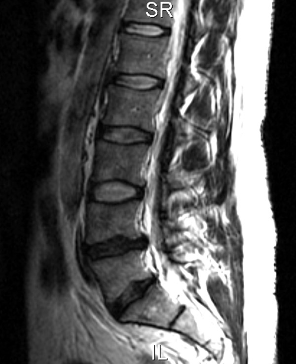
T2 weighted sagittal MRI demonstrating a loss of normal height and signal involving the L4–L5 disc and a broad-based paracentral disc bulge that contacted the thecal sac (arrow). L5-S1 disc degeneration was also present.
T2 weighted sagittal MRI demonstrating a loss of normal height and signal involving the L4–L5 disc and a broad-based paracentral disc bulge that contacted the thecal sac (arrow). L5-S1 disc degeneration was also present.