|
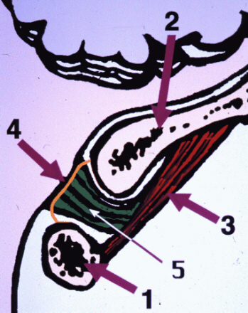
Visualization of the Muscle-Dural Bridge |
|
We have described (1) a previously unreported anatomical connection between a deep
occipital-cervical muscle, the rectus capitis posterior minor and
the dura mater, observed in 15 cadaveric specimens, and clinical
MRI scans (Figure 1)
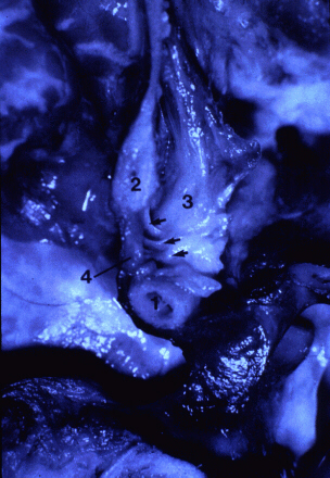
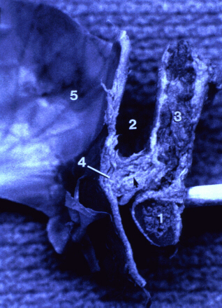
A connective tissue bridge between the rectus capitis posterior minor muscle and the dorsal
aspect of the spinal dura mater at the atlanto-occipital junction
was observed in cadaver dissections of fresh (Figure
2) and fixed (Figure 3) specimens. The fibers of the
muscle-dural bridge were oriented primarily perpendicular to the
dura. This arrangement of fibers appears to resist movement of
the dura toward the spinal cord.
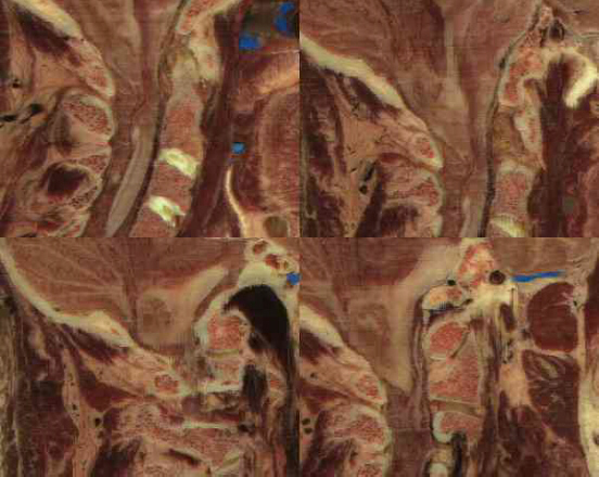
Using EAI's visualization
technology it has been possible to identify this structure in the
Visible Human Female data set. We have reformatted the data set
by reslicing it first in the coronal and the sagittal planes. We
have also reformatted the data set in five more, non-conventional
planes, between the sagittal and coronal planes, at 15 degrees
increments (Figure 4). These planes afford a better visualization of
the muscle-dural bridge than the conventional, sagittal and
coronal planes. As a general observation, most structures are
best identified when the sectional plane is either perpendicular
or parallel to the long axis of the respective structure.
Therefore, we expected to gain a better view of the muscle-dural
bridge in planes parallel to the rectus capitis posterior minor
muscle. Indeed, in the planes that are offset by an angle of 60
to 75 degrees from the coronal plane not only the RCPM is clearly
visible, along with its attachment on the atlas, but also the
connective tissue superior to the muscle, which clearly appears
to protrude between the occipital bone and the atlas into the
spinal canal. In figure 4, we have also noticed muscle fibers
that do not follow the direction of the RCPM, but seem to run
along the connective tissue fibers (second image,
clockwise).
It has been speculated that the
function of the muscle dural bridge may be to prevent folding of
the dura mater during hyperextension of the neck. Also, clinical
evidence suggests that the muscle dural bridge may play an
important role the pathogenesis of the cervicogenic headaches.
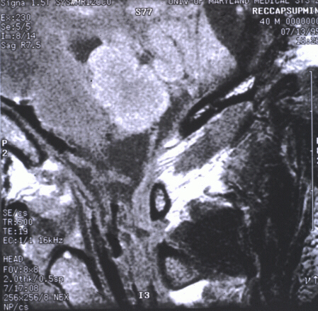
The functional research of the
muscle-dural bridge poses obvious difficulties, due to various
reasons. Its size makes it difficult to attempt functional MR
studies, although the structure has been identified in clinical
MR images (Figure 5). Therefore, we are planning to perform a
computer aided biomechanical simulation of this structure, which
is likely to yield important data regarding its
function.
Reference:
1. Hack, G.D., Koritzer, R.T.,
Robinson, W.L., Hallgren, R.C., Greenman, P.E.
Anatomic Relation
Between the Rectus Capitis Posterior Minor Muscle and the Dura Mater
Spine 1995 (Dec); 20 (23): 2484-2486
