

Spinous Process Hypertrophy Associated with
Implanted Devices in the External Link ModelThis section is compiled by Frank M. Painter, D.C.
Send all comments or additions to: Frankp@chiro.org


FROM: J Manipulative Physiol Ther 2012 (Jun); 35 (5): 367–371 ~ FULL TEXT
Nicole M. Homb, DC, Charles N.R. Henderson, DC
Palmer College of Chiropractic,
Davenport, IA, USA
OBJECTIVE: Recent development of a chiropractic subluxation mimic, the external link model, uses titanium implants on lumbar vertebrae in the rat. The objective of this study was to evaluate potential correlations in the model between linking history, bone resorption, exudate formation, and experimentally induced intervertebral hypomobility.
METHODS: Serial lateral radiographs of 73 male Sprague Dawley rats with implanted devices were reviewed. A baseline radiograph was obtained after a 6-week surgical recovery period, and a second radiograph was exposed after an 8-week hypomobility induction period. Spinous hypertrophy at the implant sites (L4, L5, and L6) was measured on the radiographs with a vernier caliper. Bone resorption and exudate build-up were assessed and compared with intervertebral hypomobility data previously collected. Data trends were described using cross-tabulated counts, analysis of variance, and regression analysis.
RESULTS: Cross-tabulation suggested differences between hypomobility-induced rats and control rats. However, correlation analysis showed no predictive role for spinous hypertrophy relative to intervertebral mobility. Similarly, exudate level did not predict spinous hypertrophy. However, implant presence and vertebral level had a significant interaction, with moderate and severe hypertrophy occurring more frequently at L4 and L6 in hypomobility-induced rats. Age did not materially influence spinous hypertrophy.
CONCLUSIONS: Mechanical stresses produced at the implant bone interface in rats with induced hypomobility contribute to spinous hypertrophy beyond that simply due to the presence of the implants. However, spinous hypertrophy does not contribute significantly to intervertebral hypomobility in the external link model.
From the FULL TEXT Article
Introduction
The external link model (ELM) was developed as a research platform to study the biologic effects of chronic intervertebral hypomobility. It is a foundational concept among practitioners of spinal manipulation that intervertebral hypomobility produces meaningful biologic effects. [1, 2] Spinal manipulative therapy is thought to increase or restore mobility to the spine. [3, 4] The ELM uses surgically implanted titanium spinous attachment units (SAUs) on the spinous processes of 3 contiguous lumbar segments (L4, L5, and L6) in the rat (Figure 1). Titanium has become the material of choice for metal implants because it is twice as strong as steel with only half the weight and is also more biocompatible. [5-7] The total weight of the 3 SAUs with links, screws, and nuts is only 1.67 g. This implant design allows investigators to induce intervertebral hypomobility and misalignment via a reversible external link system. [8, 9] Studies with this model have shown progressive increased spine stiffness (intervertebral hypomobility), [9] zygapophysial joint degeneration, [10] intra-articular (zygapophysial) adhesions, [11] and synapse morphology changes in the superficial dorsal horn of the segmentally related spinal cord. [12]
It is possible that interaction between the implant material and local tissues may also produce bone remodeling. We were interested in the possibility that spinous hypertrophy associated with the implant devices may be a significant contributor to the observed biomechanical effects. Therefore, this study examined the extent of spinous hypertrophy and its possible correlation with intervertebral mobility. To the authors' knowledge, this is the first report describing hypertrophy of spinous processes associated with implanted devices. Two clinically relevant conditions of spinous hypertrophy are reported in the literature. Baastrup's disease is a condition in which hypertrophied lumbar spinous processes approximate one another producing reactive bone sclerosis, adventitious bursae, and low back pain. [13, 14] In the Morbus de Anquin syndrome (spinous engagement syndrome), a hypertrophied lumbar spinous process combines with a spina bifida occulta resulting in cord compression and low back pain. [15] Rats bearing the implanted spinous attachment devices used in this study do not show general chronic pain behaviors (eg, decreased eating or drinking, increased resting, excessive grooming, scratching, or biting). [9, 16] However, we were concerned that approximating spinous processes due to hypertrophy might explain the intervertebral hypomobility shown in the ELM.
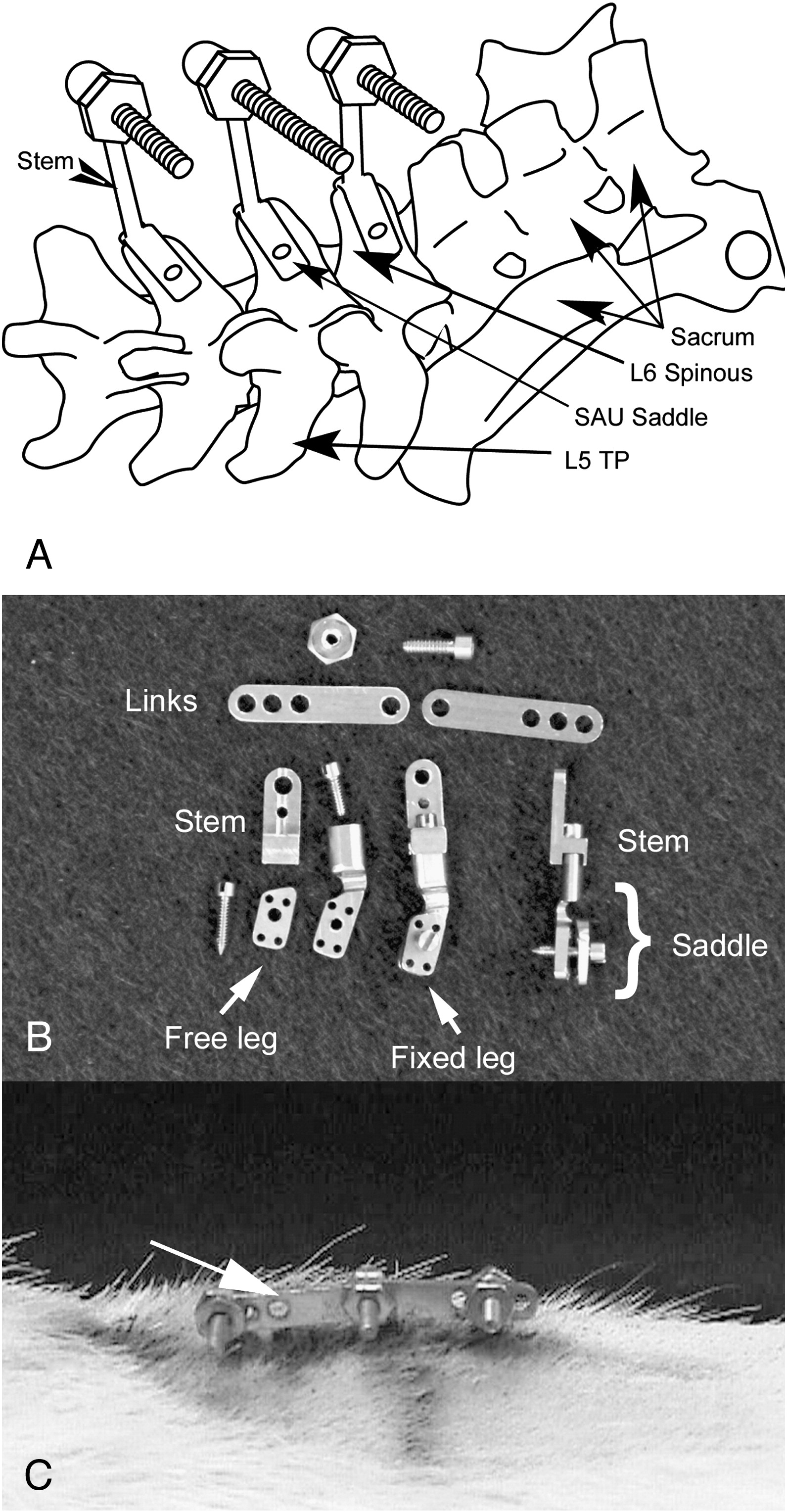
Figure 1. Spinous attachment units.
A: Superior oblique view showing placement of the L4, L5, and L6 SAUs.
The rat has 6 lumbar vertebrae.
B: Titanium, 3-piece SAUs. Each SAU has a removable stem, a saddle with a
fixed leg, and a free leg. The far right SAU is rotated to show the saddle legs
in profile, and the far left SAU is “exploded” to show the component parts.
C: Lateral view of a rat linked in the neutral position.
Hypomobility was induced by linking rats for 8 weeks.
Methods
Serial radiographs from 73 rats with implanted SAUs were examined for evidence of spinous hypertrophy. Hypertrophy data were compared with intervertebral mobility measurements obtained on the same animals. Spinous hypertrophic changes were compared between never-linked control rats (CLINK, n = 24) and rats that had SAUs linked to induce intervertebral hypomobility (ELINK, n = 49). This allowed us to distinguish between effects produced simply by the presence of SAUs on spinous processes and effects produced by linking adjacent SAUs. We report here a post hoc analysis of data collected from 2 separate studies previously approved by the Palmer College Institutional Animal Care and Use Committee. CLINK data were drawn from a National Institutes of Health/National Center for Complementary and Alternative Medicine (NIH/NCCAM)–funded study (grant no. R21 AT00784-02, Evaluating Reversible Spinal Fixation in Biped Rats) that was designed to develop and evaluate a novel bipedal rat model of the subluxation. ELINK data were drawn from another NIH/NCCAM–funded study (grant no. 1U19AT004663.01, Examining Manipulation with a Spine Fixation Model) that was designed to evaluate the behavioral, biomechanical, and biologic effects of spinal manipulation in the ELM. In these 2 studies, SAUs were implanted by researchers skilled in these implant procedures (>300 rats implanted), and the radiographs were obtained at equivalent periods.
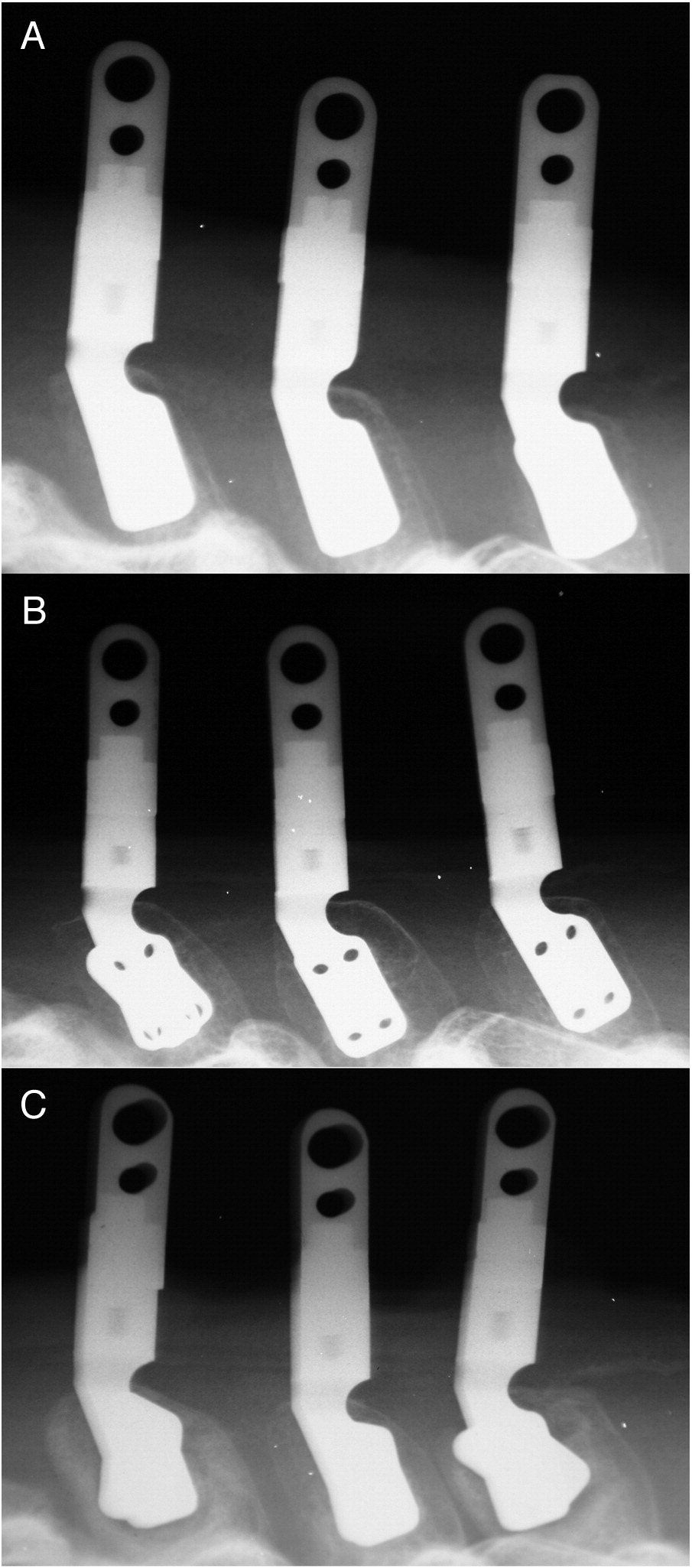
Spinous process width measurements were obtained from films exposed in a shielded x-ray cabinet (Faxitron MX-20, Faxitron X-Ray, Lincolnshire, IL) using a molded positioning jig that held the animals in a standard extended position (Figure 2). This unit's 15-µm focal spot produced very high–image resolution films (20 line pairs/mm) with a 2-fold image magnification. Initial (baseline) radiographs were obtained after a 6-week surgical recovery period with subsequent radiographs taken after an 8-week link period (or its time equivalent in control rats). The degree of hypertrophy at the L4, L5, and L6 spinous processes was determined by measuring spinous widths on radiographs with a vernier caliper (Empire, Norfolk, Virginia) using a scale (Table 1). All width measurements were taken at the approximate midlength of each spinous process (±0.25 mm precision). The mean of measurements obtained from 3 independent reviewers was calculated for each film. Bone resorption was determined by one of the authors (CNRH) using a 4-point rating scale (0-3, none-severe). The hallmark of “severe” bone resorption, grade “3,” was a complete zone of radiographic bone loss around the implant with one or more radial fracture lines in the shell of bone surrounding the lytic area. In grade “2” resorption, bone loss was clearly present but did not attain the hallmark signs of grade 3. Grade “1” bone resorption indicated that there was some suggestion of bone loss, but it was not unequivocally present. Exudate was also graded using a 4-point rating scale (0-3). Quantity of exudate was reported, as it was observed to cover the 8-mm implant stem (Figure 1): Grade 0 exudate covered less than one-third of the stem, grade 1 covered at least one-third of the stem but less than two-thirds of the stem, grade 2 covered more than two-thirds of the stem but less than the entire stem, and grade 3 covered the entire height of the stem. Figure 3 illustrates varying degrees of spinous hypertrophy on 3 radiographs. Grade 2 bone resorption may also be seen at L4 and L6 in this figure (Figure 3C).
Figure 2. Rat positioning jig.
This simple molded positioning jig held each rat in a standard extended position
during x-ray exposure. An anesthetized rat would be placed into the mold
immediately before the x-ray was exposed.

Table 1. Spinous process hypertrophy rating scale
a Corrected for the 2× radiograph magnification factor.
Rating Description Spinous width on x-ray (mm) Osseous spinous width (mm) a 0 None ≤11 ≤5.5 1 Mild (tentative hypertrophy) 11.1-13 5.6-6.5 2 Moderate 13.1-15 6.6-7.5 3 Severe >15 >7.5
Figure 3. Spinous process hypertrophy.
Spinous processes L4, L5, and L6 are shown in each of the 3 radiographs, left to right, respectively.
A: Normal spinous process width.
B: L5 spinous has moderate hypertrophy, whereas the L4 and L6 spinouses demonstrate the upper limit of mild hypertrophy.
C: All 3 spinouses show severe hypertrophy with grade 2 bone resorption at L4 and L6.
Data Analysis

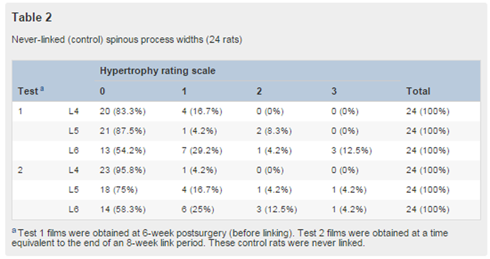
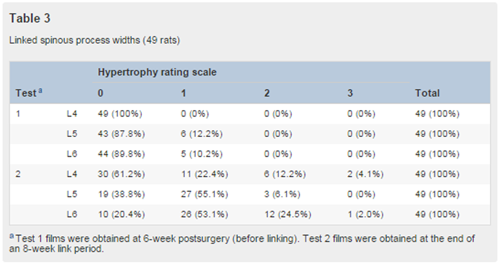
Table 2
Table 3 All analyses were performed with SPSS 15.0 for Windows (International Business Machines Corp, Armonk, NY). Spinous hypertrophy prevalence was shown by cross-tabulation for never-linked control rats and rats linked for 8 weeks (Table 2, Table 3, respectively). Linking effect, age (never-linked equivalent period), and vertebral level effects on spinous hypertrophy and bone resorption were evaluated via repeated-measures analysis of variance. Lastly, spinous hypertrophy was evaluated as a predictor of intervertebral hypomobility, and exudate production was examined as a predictor of spinous hypertrophy. A linear regression analysis was performed for these 2 relationships at each of the 2 test periods in the linked rats study group.
Results
Cross-tabulation of spinous hypertrophy prevalence in never-linked rats and linked rats (Table 2, Table 3, respectively) suggested a difference in hypertrophy prevalence between these 2 study groups. Spinous hypertrophy in the never-linked group did not change significantly over the 8-week interval between tests 1 and 2, whereas the linked group had a substantial change at all vertebral levels. In the linked group, moderate and severe hypertrophy during test 2 was seen most frequently at L4 and L6. In the 2 studies, test 1 (prelink or control time equivalent) prevalence was comparable. There were few occurrences of moderate to severe hypertrophy in test 1 (6/24 never-linked rats and 0/49 linked rats). Spinous hypertrophy did not predict changes in intervertebral motion at any segmental level (r2 = 0.08; L4, P = .76; L5, P = .12; and L6, P = .92), and exudate level did not predict the presence or severity of spinous hypertrophy (P = .71).
As suggested in the cross-tabulation tables (Table 2, Table 3); SAU linking and vertebral level had a significant interaction (analysis of variance, P = .01), with moderate and severe hypertrophy occurring more frequently at L4 and L6 after linking. Time-related (age effect) hypertrophy was not observed in control rats. An equivalent 8-week period without linking produced no significant effect on hypertrophy (P = .89).
Discussion
Results from these secondary analyses suggest that mechanical stresses put upon the implant bone interface by the external linking of the SAUs contribute to spinous hypertrophy beyond the minimal influence associated with the presence of the SAU on the bone. However, the correlation analysis shown that spinous hypertrophy does not contribute significantly to intervertebral hypomobility produced in the ELM. This is a critical finding. If spinous hypertrophy was a substantial contributor to the stiffness and hypomobility produced in the ELM, it would seriously undermine the credibility of this model as a possible subluxation mimic. It is reasonable that the L4 and L6 vertebrae show a greater frequency of moderate to severe hypertrophy in linked rats because these vertebrae should receive greater mechanical stress from adjacent mobile segments. By contrast, the L4 and L6 vertebrae would shield L5 from mechanical loading by these mobile vertebrae. Micromovement at the implant bone interface has also been shown to contribute to aseptic loosening of implants. [17, 18] This may be important in future studies examining SAU stability and the effects of longer link periods in the ELM.
We were surprised to find that exudate levels did not correlate with spinous hypertrophy or bone resorption. Recent studies suggest that bone resorption around implants and exudate production may be due to endotoxins that are killed bacteria residues. [19, 20] Consequently, the ELM protocol includes a rigorous cleaning procedure for reused SAUs to prevent bacteria residue induced periprosthetic osteolysis. [21]
Study Limitations
Although the findings of these secondary analyses are informative, we acknowledge the interpretive limitations associated with post hoc analyses. The research questions examined in this study were not specifically addressed in the objectives and design of the original studies from which these data arise. However, these findings do provide important guidance for future, a priori investigations into issues of spinous hypertrophy, bone resorption, and intervertebral hypomobility in the ELM. The ELM is a novel research platform in that it permits examination of long-term effects associated with intervertebral mobility. [8] Further studies are necessary to better define both its strengths and its limitations.
Conclusion
Results from these secondary analyses suggest that mechanical stresses produced on the implant bone interface by links in the ELM contribute to spinous hypertrophy beyond those associated with the presence of the SAU on the bone. In addition, the study findings suggest that spinous hypertrophy does not contribute significantly to intervertebral hypomobility produced in the ELM. This is important because it supports the argument that stiffness and hypomobility produced in the ELM are not materially an artifact of spinous hypertrophy. The analysis upon which these findings were drawn is a secondary analysis of data drawn from studies not specifically designed to address these research questions. Consequently, this study does not validate the ELM as a research tool; but it does illuminate the path for future ELM investigations.
Funding Sources and Potential Conflicts of Interest
The work reported in this manuscript was supported by NIH/NCCAM grants 1U19AT004663.01 and R21 AT00784-02 . The research was conducted in a facility constructed with support from Research Facilities Improvement Grant C06 RR15433 from the National Center for Research Resources, National Institutes of Health. No conflicts of interest were reported for this study.
Practical Applications
Spinous hypertrophy does not contribute significantly to induced intervertebral hypomobility in the ELM.
This finding strengthens the argument that the ELM is a useful experimental platform for studying the chiropractic subluxation.
References:
Gatterman, M.I.
Foundations of chiropractic: subluxation. 2nd ed.
Foundations of chiropractic: subluxation. 2nd ed.
Elsevier Mosby, St. Louis; 2005Leach, R.A.
The chiropractic theories: a textbook of scientific research. 4th ed.
Lippincott Williams & Wilkins, Philadelphia; 2004: 1–463Henderson, C.N.R.
Three neurophysiologic theories on the chiropractic subluxation.
in: Foundations of chiropractic: subluxation. 2nd ed.
Elsevier Mosby, St. Louis; 2005: 296–303Pickar JG.
Neurophysiological Effects of Spinal Manipulation
Spine J (N American Spine Society) 2002 (Sep); 2 (5): 357–371Assad, M., Lemieux, N., Rivard, C.H., and Yahia, L.H.
Comparative in vitro biocompatibility of nickel-titanium, pure nickel, pure titanium,
and stainless steel: genotoxicity and atomic absorption evaluation.
Biomed Mater Eng. 1999; 9: 1–12Ysander, M., Brĺnemark, R., Olmarker, K., and Myers, R.R.
Intramedullary osseointegration: development of a rodent model and study of
histology and neuropeptide changes around titanium implants.
J Rehabil Res Dev. 2001; 38: 183–190Hallab, N., Link, H.D., and McAfee, P.C.
Biomaterial optimization in total disc arthroplasty.
Spine. 2003; 28: S139–S152Henderson, C.N., Cramer, G.D., Zhang, Q.,
DeVocht, J.W., and Fournier, J.T.
Introducing the External Link Model for Studying Spine Fixation and Misalignment: Part 1
Need, Rationale, and Applications
J Manipulative Physiol Ther 2007 (Mar); 30 (3): 239–245Henderson, C.N., Cramer, G.D., Zhang, Q.,
DeVocht, J.W., and Fournier, J.T.
Introducing the External Link Model for Studying Spine Fixation and Misalignment: Part 2
Biomechanical Features
J Manipulative Physiol Ther 2007 (May); 30 (4): 279–294Cramer G.D., Fournier J.T., Henderson C.N., Wolcott C.C.
Degenerative Changes Following Spinal Fixation in a Small Animal Model
J Manipulative Physiol Ther 2004 (Mar); 27 (3): 141–154Cramer GD, Henderson CNR, Little JW et al.
Zygapophyseal Joint Adhesions After Induced Hypomobility
J Manipulative Physiol Ther. 2010 (Sep); 33 (7): 508–518Bakkum, B.W., Henderson, C.N., Hong, S.P., and Cramer, G.D.
Preliminary Morphological Evidence That Vertebral Hypomobility Induces Synaptic Plasticity
in the Spinal Cord
J Manipulative Physiol Ther. 2007 (Jun); 30 (5): 336–342Clifford, P.D.
Baastrup disease.
Am J Orthop. 2007; 36: 560–561Pinto, P.S., Boutin, R.D., and Resnick, D.
Spinous process fractures associated with Baastrup disease.
Clin Imaging. 2004; 28: 219–222Bruns, J., Rehder, U., Dahmen, G.P., Behrens, P., and Meiss, L.
Morbus de Anquin or spinous engagement syndrome. A rare cause of low-back pain syndrome and sciatica.
Eur Spine J. 1994; 3: 265–269Whishaw, I.Q. and Kolb, B.
The behavior of the laboratory rat a handbook with tests.
Oxford University Press, Oxford; 2005Jones, L.C., Frondoza, C., and Hungerford, D.S.
Effect of PMMA particles and movement on an implant interface in a canine model.
J Bone Joint Surg Br. 2001; 83: 448–458Jasty, M., Bragdon, C., Burke, D., O'Connor, D.,
Lowenstein, J., and Harris, W.H.
In vivo skeletal responses to porous-surfaced implants subjected to small induced motions.
J Bone Joint Surg Am. 1997; 79: 707–714Sundfeldt, M., Carlsson, L.V., Johansson, C.B.,
Thomsen, P., and Gretzer, C.
Aseptic loosening, not only a question of wear: a review of different theories.
Acta Orthop. 2006; 77: 177–197Greenfield, E.M., Bi, Y., Ragab, A.A.,
Goldberg, V.M., Nalepka, J.L., and Seabold, J.M.
Does endotoxin contribute to aseptic loosening of orthopedic implants?.
J Biomed Mater Res B Appl Biomater. 2005; 72: 79–85Henderson, C. N., Cramer, G. D., Zhang, Q., DeVocht, J. W.,
Sozio, R. S., & Fournier, J. T.
Introducing the External Link Model for Studying Spine Fixation and Misalignment:
Current Procedures, Costs, and Failure Rates
J Manipulative Physiol Ther 2009 (May); 32 (4): 294–302
Return to SUBLUXATION DEGENERATION
Since 2-25-2016
| Home Page | Visit Our Sponsors | Become a Sponsor |
Please read our DISCLAIMER |